Li Zhang, Kaidong Chen, Yan Zhi, Wei Ji, Guofeng Shi, Yiping You, Feng Wang, Kefei Chen, Tian Xu, Xiangming Fang
{"title":"帕金森病患者脑铁沉积与纯粹冷漠之间的关系:一项横断面定量易感性成像研究","authors":"Li Zhang, Kaidong Chen, Yan Zhi, Wei Ji, Guofeng Shi, Yiping You, Feng Wang, Kefei Chen, Tian Xu, Xiangming Fang","doi":"10.21037/qims-2024-2626","DOIUrl":null,"url":null,"abstract":"<p><strong>Background: </strong>Apathy, a decline in goal-directed motivated behavior, is a common non-motor symptom (NMS) in Parkinson's disease (PD). Previous studies have suggested that PD patients with apathy exhibit increased iron levels in the cerebrospinal fluid (CSF) and the iron levels are positively correlated with the severity of apathy, indicating that apathy in PD may be related with brain iron accumulation. Specifically, quantitative susceptibility mapping (QSM), an emerging brain magnetic resonance imaging (MRI) technique, can be used to sensitively detect the iron deposition in the brain <i>in vivo</i>, to reflect the neurodegeneration processes. This study thus used QSM to detect the brain iron deposition in PD with pure apathy (PD-PA) and examined apathy symptoms-related regional brain iron deposition, and aimed to explore the underlying mechanism of neurodegeneration of PD-PA.</p><p><strong>Methods: </strong>A total of 29 patients with PD-PA, 33 PD without pure apathy (PD-NPA), and 32 healthy controls (HCs) were collected. All participants underwent three-dimensional T1-weighted imaging (3DT1) and QSM scans. The susceptibility values of PD-PA, PD-NPA, and HC groups were compared at whole brain voxel-wise level and the region of interest (ROI)-wise level, respectively. Moreover, correlation analysis between apathy symptoms and brain iron deposition was further performed in the PD-PA group.</p><p><strong>Results: </strong>Relative to the HCs, patients with PD demonstrated increased susceptibility values (consistent with higher brain tissue iron deposits) in substantia nigra pars compacta (SNc) and ventral tegmental area (VTA) of the midbrain [P<sub>false discovery rate (FDR)</sub><0.05]. Further PD subgroup analysis suggested that compared with HCs, patients with PD-NPA showed increased iron deposition in the right medial superior frontal gyrus (SFGmed), whereas those with PD-PA exhibited more extensive increased iron deposition in SFGmed, extending to bilateral SFGmed [voxel-level P<0.001, cluster-level P<sub>family-wise error (FWE)</sub><0.05]. The Apathy Scale (AS) score was positively correlated with the mean susceptibility values of the left SFGmed in the PD-PA group (r=0.651, P=0.001). In the PD-PA group, voxel-wise whole-brain correlation analysis found a positive correlation between the AS score and the susceptibility values in the left SFGmed, left anterior cingulate cortex (ACC), right thalamus, as well as the right superior temporal gyrus (STG) (voxel-level P<0.001, cluster-level P<sub>FWE</sub><0.05).</p><p><strong>Conclusions: </strong>In patients with PD-PA, brain regions associated with brain iron deposition were mainly located in the \"mesocorticolimbic loop\" (SFGmed and ACC), as well as the STG and thalamus. The present study suggested that abnormal iron deposition in these core brain regions may be associated with abnormal top-down and bottom-up neuroregulation and involved in the mechanism of PD apathy. The present study provides new insights into the pathophysiologic mechanism of PD-PA.</p>","PeriodicalId":54267,"journal":{"name":"Quantitative Imaging in Medicine and Surgery","volume":"15 9","pages":"8292-8308"},"PeriodicalIF":2.3000,"publicationDate":"2025-09-01","publicationTypes":"Journal Article","fieldsOfStudy":null,"isOpenAccess":false,"openAccessPdf":"https://www.ncbi.nlm.nih.gov/pmc/articles/PMC12397670/pdf/","citationCount":"0","resultStr":"{\"title\":\"Association between brain iron deposition and pure apathy in Parkinson's disease: a cross-sectional quantitative susceptibility mapping imaging study.\",\"authors\":\"Li Zhang, Kaidong Chen, Yan Zhi, Wei Ji, Guofeng Shi, Yiping You, Feng Wang, Kefei Chen, Tian Xu, Xiangming Fang\",\"doi\":\"10.21037/qims-2024-2626\",\"DOIUrl\":null,\"url\":null,\"abstract\":\"<p><strong>Background: </strong>Apathy, a decline in goal-directed motivated behavior, is a common non-motor symptom (NMS) in Parkinson's disease (PD). Previous studies have suggested that PD patients with apathy exhibit increased iron levels in the cerebrospinal fluid (CSF) and the iron levels are positively correlated with the severity of apathy, indicating that apathy in PD may be related with brain iron accumulation. Specifically, quantitative susceptibility mapping (QSM), an emerging brain magnetic resonance imaging (MRI) technique, can be used to sensitively detect the iron deposition in the brain <i>in vivo</i>, to reflect the neurodegeneration processes. This study thus used QSM to detect the brain iron deposition in PD with pure apathy (PD-PA) and examined apathy symptoms-related regional brain iron deposition, and aimed to explore the underlying mechanism of neurodegeneration of PD-PA.</p><p><strong>Methods: </strong>A total of 29 patients with PD-PA, 33 PD without pure apathy (PD-NPA), and 32 healthy controls (HCs) were collected. All participants underwent three-dimensional T1-weighted imaging (3DT1) and QSM scans. The susceptibility values of PD-PA, PD-NPA, and HC groups were compared at whole brain voxel-wise level and the region of interest (ROI)-wise level, respectively. Moreover, correlation analysis between apathy symptoms and brain iron deposition was further performed in the PD-PA group.</p><p><strong>Results: </strong>Relative to the HCs, patients with PD demonstrated increased susceptibility values (consistent with higher brain tissue iron deposits) in substantia nigra pars compacta (SNc) and ventral tegmental area (VTA) of the midbrain [P<sub>false discovery rate (FDR)</sub><0.05]. Further PD subgroup analysis suggested that compared with HCs, patients with PD-NPA showed increased iron deposition in the right medial superior frontal gyrus (SFGmed), whereas those with PD-PA exhibited more extensive increased iron deposition in SFGmed, extending to bilateral SFGmed [voxel-level P<0.001, cluster-level P<sub>family-wise error (FWE)</sub><0.05]. The Apathy Scale (AS) score was positively correlated with the mean susceptibility values of the left SFGmed in the PD-PA group (r=0.651, P=0.001). In the PD-PA group, voxel-wise whole-brain correlation analysis found a positive correlation between the AS score and the susceptibility values in the left SFGmed, left anterior cingulate cortex (ACC), right thalamus, as well as the right superior temporal gyrus (STG) (voxel-level P<0.001, cluster-level P<sub>FWE</sub><0.05).</p><p><strong>Conclusions: </strong>In patients with PD-PA, brain regions associated with brain iron deposition were mainly located in the \\\"mesocorticolimbic loop\\\" (SFGmed and ACC), as well as the STG and thalamus. The present study suggested that abnormal iron deposition in these core brain regions may be associated with abnormal top-down and bottom-up neuroregulation and involved in the mechanism of PD apathy. The present study provides new insights into the pathophysiologic mechanism of PD-PA.</p>\",\"PeriodicalId\":54267,\"journal\":{\"name\":\"Quantitative Imaging in Medicine and Surgery\",\"volume\":\"15 9\",\"pages\":\"8292-8308\"},\"PeriodicalIF\":2.3000,\"publicationDate\":\"2025-09-01\",\"publicationTypes\":\"Journal Article\",\"fieldsOfStudy\":null,\"isOpenAccess\":false,\"openAccessPdf\":\"https://www.ncbi.nlm.nih.gov/pmc/articles/PMC12397670/pdf/\",\"citationCount\":\"0\",\"resultStr\":null,\"platform\":\"Semanticscholar\",\"paperid\":null,\"PeriodicalName\":\"Quantitative Imaging in Medicine and Surgery\",\"FirstCategoryId\":\"3\",\"ListUrlMain\":\"https://doi.org/10.21037/qims-2024-2626\",\"RegionNum\":2,\"RegionCategory\":\"医学\",\"ArticlePicture\":[],\"TitleCN\":null,\"AbstractTextCN\":null,\"PMCID\":null,\"EPubDate\":\"2025/8/12 0:00:00\",\"PubModel\":\"Epub\",\"JCR\":\"Q2\",\"JCRName\":\"RADIOLOGY, NUCLEAR MEDICINE & MEDICAL IMAGING\",\"Score\":null,\"Total\":0}","platform":"Semanticscholar","paperid":null,"PeriodicalName":"Quantitative Imaging in Medicine and Surgery","FirstCategoryId":"3","ListUrlMain":"https://doi.org/10.21037/qims-2024-2626","RegionNum":2,"RegionCategory":"医学","ArticlePicture":[],"TitleCN":null,"AbstractTextCN":null,"PMCID":null,"EPubDate":"2025/8/12 0:00:00","PubModel":"Epub","JCR":"Q2","JCRName":"RADIOLOGY, NUCLEAR MEDICINE & MEDICAL IMAGING","Score":null,"Total":0}
Association between brain iron deposition and pure apathy in Parkinson's disease: a cross-sectional quantitative susceptibility mapping imaging study.
Background: Apathy, a decline in goal-directed motivated behavior, is a common non-motor symptom (NMS) in Parkinson's disease (PD). Previous studies have suggested that PD patients with apathy exhibit increased iron levels in the cerebrospinal fluid (CSF) and the iron levels are positively correlated with the severity of apathy, indicating that apathy in PD may be related with brain iron accumulation. Specifically, quantitative susceptibility mapping (QSM), an emerging brain magnetic resonance imaging (MRI) technique, can be used to sensitively detect the iron deposition in the brain in vivo, to reflect the neurodegeneration processes. This study thus used QSM to detect the brain iron deposition in PD with pure apathy (PD-PA) and examined apathy symptoms-related regional brain iron deposition, and aimed to explore the underlying mechanism of neurodegeneration of PD-PA.
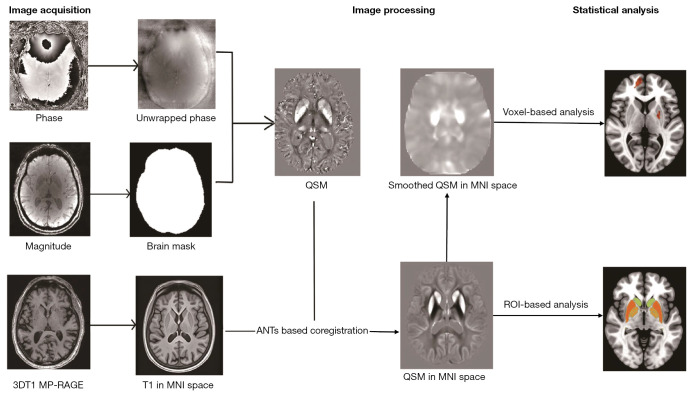
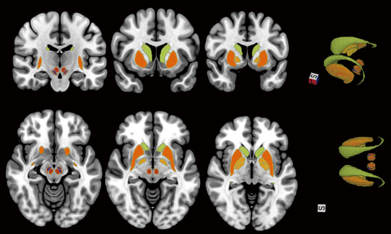
Methods: A total of 29 patients with PD-PA, 33 PD without pure apathy (PD-NPA), and 32 healthy controls (HCs) were collected. All participants underwent three-dimensional T1-weighted imaging (3DT1) and QSM scans. The susceptibility values of PD-PA, PD-NPA, and HC groups were compared at whole brain voxel-wise level and the region of interest (ROI)-wise level, respectively. Moreover, correlation analysis between apathy symptoms and brain iron deposition was further performed in the PD-PA group.
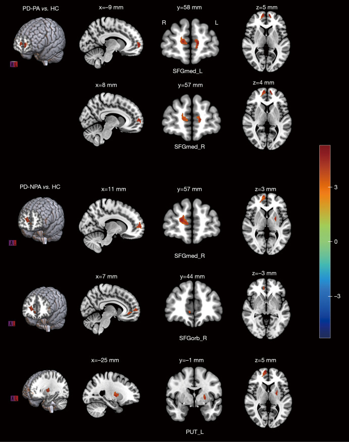
Results: Relative to the HCs, patients with PD demonstrated increased susceptibility values (consistent with higher brain tissue iron deposits) in substantia nigra pars compacta (SNc) and ventral tegmental area (VTA) of the midbrain [Pfalse discovery rate (FDR)<0.05]. Further PD subgroup analysis suggested that compared with HCs, patients with PD-NPA showed increased iron deposition in the right medial superior frontal gyrus (SFGmed), whereas those with PD-PA exhibited more extensive increased iron deposition in SFGmed, extending to bilateral SFGmed [voxel-level P<0.001, cluster-level Pfamily-wise error (FWE)<0.05]. The Apathy Scale (AS) score was positively correlated with the mean susceptibility values of the left SFGmed in the PD-PA group (r=0.651, P=0.001). In the PD-PA group, voxel-wise whole-brain correlation analysis found a positive correlation between the AS score and the susceptibility values in the left SFGmed, left anterior cingulate cortex (ACC), right thalamus, as well as the right superior temporal gyrus (STG) (voxel-level P<0.001, cluster-level PFWE<0.05).
Conclusions: In patients with PD-PA, brain regions associated with brain iron deposition were mainly located in the "mesocorticolimbic loop" (SFGmed and ACC), as well as the STG and thalamus. The present study suggested that abnormal iron deposition in these core brain regions may be associated with abnormal top-down and bottom-up neuroregulation and involved in the mechanism of PD apathy. The present study provides new insights into the pathophysiologic mechanism of PD-PA.

 求助内容:
求助内容: 应助结果提醒方式:


