Xiaochen Feng, Yaying Zhang, Minming Lu, Chao Ma, Xiaoqiang Miao, Jiacheng Yang, Lina Lin, Yueyi Zhang, Kai Zhang, Ning Zhang, Yan Kang, Yu Luo, Kai Cao
{"title":"基于深度学习的MRI全自动评估颈椎管狭窄的可行性。","authors":"Xiaochen Feng, Yaying Zhang, Minming Lu, Chao Ma, Xiaoqiang Miao, Jiacheng Yang, Lina Lin, Yueyi Zhang, Kai Zhang, Ning Zhang, Yan Kang, Yu Luo, Kai Cao","doi":"10.21037/qims-2025-67","DOIUrl":null,"url":null,"abstract":"<p><strong>Background: </strong>Currently, there is no fully automated tool available for evaluating the degree of cervical spinal stenosis. The aim of this study was to develop and validate the use of artificial intelligence (AI) algorithms for the assessment of cervical spinal stenosis.</p><p><strong>Methods: </strong>In this retrospective multi-center study, cervical spine magnetic resonance imaging (MRI) scans obtained from July 2020 to June 2023 were included. Studies of patients with spinal instrumentation or studies with suboptimal image quality were excluded. Sagittal T2-weighted images were used. The training data from the Fourth People's Hospital of Shanghai (Hos. 1) and Shanghai Changzheng Hospital (Hos. 2) were annotated by two musculoskeletal (MSK) radiologists following Kang's system as the standard reference. First, a convolutional neural network (CNN) was trained to detect the region of interest (ROI), with a second Transformer for classification. The performance of the deep learning (DL) model was assessed on an internal test set from Hos. 2 and an external test set from Shanghai Changhai Hospital (Hos. 3), and compared among six readers. Metrics such as detection precision, interrater agreement, sensitivity (SEN), and specificity (SPE) were calculated.</p><p><strong>Results: </strong>Overall, 795 patients were analyzed (mean age ± standard deviation, 55±14 years; 346 female), with 589 in the training (75%) and validation (25%) sets, 206 in the internal test set, and 95 in the external test set. Four tasks with different clinical application scenarios were trained, and their accuracy (ACC) ranged from 0.8993 to 0.9532. When using a Kang system score of ≥2 as a threshold for diagnosing central cervical canal stenosis in the internal test set, both the algorithm and six readers achieved similar areas under the receiver operating characteristic curve (AUCs) of 0.936 [95% confidence interval (CI): 0.916-0.955], with a SEN of 90.3% and SPE of 93.8%; the AUC of the DL model was 0.931 (95% CI: 0.917-0.946), with a SEN in the external test set of 100%, and a SPE of 86.3%. Correlation analysis comparing the DL method, the six readers, and MRI reports between the reference standard showed a moderate correlation, with R values ranging from 0.589 to 0.668. The DL model produced approximately the same upgrades (9.2%) and downgrades (5.1%) as the six readers.</p><p><strong>Conclusions: </strong>The DL model could fully automatically and reliably assess cervical canal stenosis using MRI scans.</p>","PeriodicalId":54267,"journal":{"name":"Quantitative Imaging in Medicine and Surgery","volume":"15 9","pages":"8457-8470"},"PeriodicalIF":2.3000,"publicationDate":"2025-09-01","publicationTypes":"Journal Article","fieldsOfStudy":null,"isOpenAccess":false,"openAccessPdf":"https://www.ncbi.nlm.nih.gov/pmc/articles/PMC12397646/pdf/","citationCount":"0","resultStr":"{\"title\":\"Feasibility of fully automatic assessment of cervical canal stenosis using MRI via deep learning.\",\"authors\":\"Xiaochen Feng, Yaying Zhang, Minming Lu, Chao Ma, Xiaoqiang Miao, Jiacheng Yang, Lina Lin, Yueyi Zhang, Kai Zhang, Ning Zhang, Yan Kang, Yu Luo, Kai Cao\",\"doi\":\"10.21037/qims-2025-67\",\"DOIUrl\":null,\"url\":null,\"abstract\":\"<p><strong>Background: </strong>Currently, there is no fully automated tool available for evaluating the degree of cervical spinal stenosis. The aim of this study was to develop and validate the use of artificial intelligence (AI) algorithms for the assessment of cervical spinal stenosis.</p><p><strong>Methods: </strong>In this retrospective multi-center study, cervical spine magnetic resonance imaging (MRI) scans obtained from July 2020 to June 2023 were included. Studies of patients with spinal instrumentation or studies with suboptimal image quality were excluded. Sagittal T2-weighted images were used. The training data from the Fourth People's Hospital of Shanghai (Hos. 1) and Shanghai Changzheng Hospital (Hos. 2) were annotated by two musculoskeletal (MSK) radiologists following Kang's system as the standard reference. First, a convolutional neural network (CNN) was trained to detect the region of interest (ROI), with a second Transformer for classification. The performance of the deep learning (DL) model was assessed on an internal test set from Hos. 2 and an external test set from Shanghai Changhai Hospital (Hos. 3), and compared among six readers. Metrics such as detection precision, interrater agreement, sensitivity (SEN), and specificity (SPE) were calculated.</p><p><strong>Results: </strong>Overall, 795 patients were analyzed (mean age ± standard deviation, 55±14 years; 346 female), with 589 in the training (75%) and validation (25%) sets, 206 in the internal test set, and 95 in the external test set. Four tasks with different clinical application scenarios were trained, and their accuracy (ACC) ranged from 0.8993 to 0.9532. When using a Kang system score of ≥2 as a threshold for diagnosing central cervical canal stenosis in the internal test set, both the algorithm and six readers achieved similar areas under the receiver operating characteristic curve (AUCs) of 0.936 [95% confidence interval (CI): 0.916-0.955], with a SEN of 90.3% and SPE of 93.8%; the AUC of the DL model was 0.931 (95% CI: 0.917-0.946), with a SEN in the external test set of 100%, and a SPE of 86.3%. Correlation analysis comparing the DL method, the six readers, and MRI reports between the reference standard showed a moderate correlation, with R values ranging from 0.589 to 0.668. The DL model produced approximately the same upgrades (9.2%) and downgrades (5.1%) as the six readers.</p><p><strong>Conclusions: </strong>The DL model could fully automatically and reliably assess cervical canal stenosis using MRI scans.</p>\",\"PeriodicalId\":54267,\"journal\":{\"name\":\"Quantitative Imaging in Medicine and Surgery\",\"volume\":\"15 9\",\"pages\":\"8457-8470\"},\"PeriodicalIF\":2.3000,\"publicationDate\":\"2025-09-01\",\"publicationTypes\":\"Journal Article\",\"fieldsOfStudy\":null,\"isOpenAccess\":false,\"openAccessPdf\":\"https://www.ncbi.nlm.nih.gov/pmc/articles/PMC12397646/pdf/\",\"citationCount\":\"0\",\"resultStr\":null,\"platform\":\"Semanticscholar\",\"paperid\":null,\"PeriodicalName\":\"Quantitative Imaging in Medicine and Surgery\",\"FirstCategoryId\":\"3\",\"ListUrlMain\":\"https://doi.org/10.21037/qims-2025-67\",\"RegionNum\":2,\"RegionCategory\":\"医学\",\"ArticlePicture\":[],\"TitleCN\":null,\"AbstractTextCN\":null,\"PMCID\":null,\"EPubDate\":\"2025/8/19 0:00:00\",\"PubModel\":\"Epub\",\"JCR\":\"Q2\",\"JCRName\":\"RADIOLOGY, NUCLEAR MEDICINE & MEDICAL IMAGING\",\"Score\":null,\"Total\":0}","platform":"Semanticscholar","paperid":null,"PeriodicalName":"Quantitative Imaging in Medicine and Surgery","FirstCategoryId":"3","ListUrlMain":"https://doi.org/10.21037/qims-2025-67","RegionNum":2,"RegionCategory":"医学","ArticlePicture":[],"TitleCN":null,"AbstractTextCN":null,"PMCID":null,"EPubDate":"2025/8/19 0:00:00","PubModel":"Epub","JCR":"Q2","JCRName":"RADIOLOGY, NUCLEAR MEDICINE & MEDICAL IMAGING","Score":null,"Total":0}
Feasibility of fully automatic assessment of cervical canal stenosis using MRI via deep learning.
Background: Currently, there is no fully automated tool available for evaluating the degree of cervical spinal stenosis. The aim of this study was to develop and validate the use of artificial intelligence (AI) algorithms for the assessment of cervical spinal stenosis.
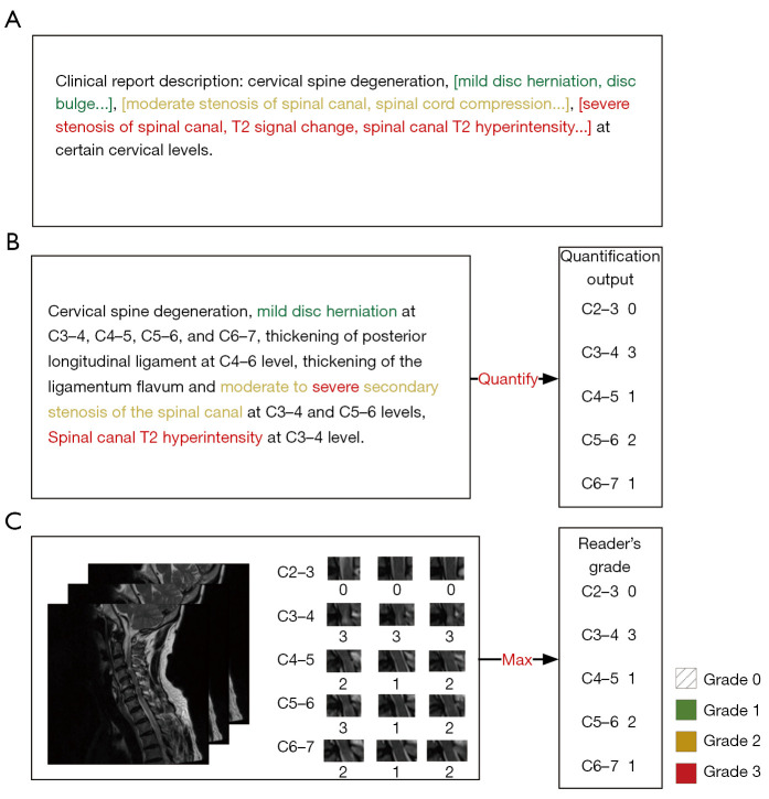
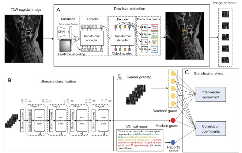
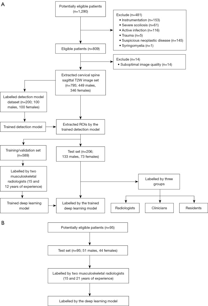
Methods: In this retrospective multi-center study, cervical spine magnetic resonance imaging (MRI) scans obtained from July 2020 to June 2023 were included. Studies of patients with spinal instrumentation or studies with suboptimal image quality were excluded. Sagittal T2-weighted images were used. The training data from the Fourth People's Hospital of Shanghai (Hos. 1) and Shanghai Changzheng Hospital (Hos. 2) were annotated by two musculoskeletal (MSK) radiologists following Kang's system as the standard reference. First, a convolutional neural network (CNN) was trained to detect the region of interest (ROI), with a second Transformer for classification. The performance of the deep learning (DL) model was assessed on an internal test set from Hos. 2 and an external test set from Shanghai Changhai Hospital (Hos. 3), and compared among six readers. Metrics such as detection precision, interrater agreement, sensitivity (SEN), and specificity (SPE) were calculated.
Results: Overall, 795 patients were analyzed (mean age ± standard deviation, 55±14 years; 346 female), with 589 in the training (75%) and validation (25%) sets, 206 in the internal test set, and 95 in the external test set. Four tasks with different clinical application scenarios were trained, and their accuracy (ACC) ranged from 0.8993 to 0.9532. When using a Kang system score of ≥2 as a threshold for diagnosing central cervical canal stenosis in the internal test set, both the algorithm and six readers achieved similar areas under the receiver operating characteristic curve (AUCs) of 0.936 [95% confidence interval (CI): 0.916-0.955], with a SEN of 90.3% and SPE of 93.8%; the AUC of the DL model was 0.931 (95% CI: 0.917-0.946), with a SEN in the external test set of 100%, and a SPE of 86.3%. Correlation analysis comparing the DL method, the six readers, and MRI reports between the reference standard showed a moderate correlation, with R values ranging from 0.589 to 0.668. The DL model produced approximately the same upgrades (9.2%) and downgrades (5.1%) as the six readers.
Conclusions: The DL model could fully automatically and reliably assess cervical canal stenosis using MRI scans.

 求助内容:
求助内容: 应助结果提醒方式:


