Deying Wen, Wen Li, Ling Zhao, Qinglin Du, Xiaoyu Tong, Ailin Liang, Tengxin Wang, Zheng Li, Xiaodi Zhang, Haiwei Liu, Yan Ren, Jiayu Sun
{"title":"肾上腺腺瘤表征的双层光谱检测器计算机断层扫描:辐射剂量减少和多相虚拟非对比成像与真实非对比成像的定量一致。","authors":"Deying Wen, Wen Li, Ling Zhao, Qinglin Du, Xiaoyu Tong, Ailin Liang, Tengxin Wang, Zheng Li, Xiaodi Zhang, Haiwei Liu, Yan Ren, Jiayu Sun","doi":"10.21037/qims-2025-854","DOIUrl":null,"url":null,"abstract":"<p><strong>Background: </strong>Computed tomography (CT) is the preferred imaging modality for evaluating adrenal lesions; however, the associated radiation exposure remains a significant concern. Dual-layer spectral detector CT (SDCT)-derived virtual noncontrast (VNC) images may reduce radiation exposure by eliminating dedicated noncontrast scans, yet their agreement with true noncontrast (TNC) imaging remains debated. This study aimed to quantitatively evaluate the agreement and image quality of VNC images [reconstructed from the arterial phase (VNCa) and portal venous phase (VNCp)] compared to TNC images in adrenal adenomas stratified by lipid content, and to assess the radiation dose reduction.</p><p><strong>Methods: </strong>A total of 103 patients with adrenal adenomas treated at the Adrenal Disease Center of West China Hospital of Sichuan University between March 2023 and September 2024 were enrolled in this prospective study. All patients underwent dual-layer SDCT examination, including TNC and arterial and venous phase scans. VNC images were reconstructed from contrast-enhanced phases. Objective metrics, including CT attenuation value [Hounsfield units (HU)], noise (standard deviation), signal-to-noise ratio (SNR), contrast-to-noise ratio, and absolute attenuation error, and subjective image quality were compared. Interobserver agreement was assessed through the calculation of interclass correlation coefficients. For objective and subjective comparisons between TNC and VNC images, statistical analyses were performed with paired <i>t</i>-tests and Wilcoxon signed-rank tests. The radiation dose with and without TNC was calculated.</p><p><strong>Results: </strong>This study included 103 patients (48 males and 55 females) with a mean age of 51.33±12.55 years. A total of 123 adrenal adenomas were identified, including 28 lipid-rich adenomas and 95 lipid-poor adenomas. For lipid-poor adenomas, VNC and TNC images showed excellent agreement in CT attenuation values (P>0.05), and compared to VNCp images, VNCa images exhibited significantly lower noise (17.44±3.39 <i>vs.</i> 18.64±2.91 HU; P<0.001) and higher SNR (1.68±0.76 <i>vs.</i> 1.55±0.67; P<0.001). In lipid-rich adenomas, VNC images overestimated CT attenuation, showing high absolute attenuation errors (VNCaerror: 9.92±6.49 HU; VNCperror: 8.50±5.17 HU), although these remained within the acceptable threshold of ≤10 HU. In the subjective scores of image quality, TNC images outperformed VNC images [TNC: median 5, interquartile range (IQR) 5-5; VNC: median 5 (IQR 4-5); P<0.001], although VNC scores remained high. No significant statistical difference was observed between the VNCa and VNCp scores (P>0.05). For most of the surrounding nonadenoma tissues, VNC and TNC images demonstrated good agreement, with attenuation differences consistently within ≤10 HU. Replacing TNC images with VNCa images could reduce the effective dose by approximately 32.63% for lipid-poor adenomas.</p><p><strong>Conclusions: </strong>Our findings suggest that for lipid-poor adenomas, VNCa demonstrates high agreement with TNC and provides superior image quality, supporting its use as a TNC substitute for reduced radiation dose. For lipid-rich adenomas, VNC should be applied with caution due to the potential risk of attenuation overestimation. Subtype classification remains essential in such studies.</p>","PeriodicalId":54267,"journal":{"name":"Quantitative Imaging in Medicine and Surgery","volume":"15 9","pages":"7935-7950"},"PeriodicalIF":2.3000,"publicationDate":"2025-09-01","publicationTypes":"Journal Article","fieldsOfStudy":null,"isOpenAccess":false,"openAccessPdf":"https://www.ncbi.nlm.nih.gov/pmc/articles/PMC12397641/pdf/","citationCount":"0","resultStr":"{\"title\":\"Dual-layer spectral detector computed tomography for adrenal adenoma characterization: radiation dose reduction and quantitative agreement of multiphase virtual noncontrast with true noncontrast imaging.\",\"authors\":\"Deying Wen, Wen Li, Ling Zhao, Qinglin Du, Xiaoyu Tong, Ailin Liang, Tengxin Wang, Zheng Li, Xiaodi Zhang, Haiwei Liu, Yan Ren, Jiayu Sun\",\"doi\":\"10.21037/qims-2025-854\",\"DOIUrl\":null,\"url\":null,\"abstract\":\"<p><strong>Background: </strong>Computed tomography (CT) is the preferred imaging modality for evaluating adrenal lesions; however, the associated radiation exposure remains a significant concern. Dual-layer spectral detector CT (SDCT)-derived virtual noncontrast (VNC) images may reduce radiation exposure by eliminating dedicated noncontrast scans, yet their agreement with true noncontrast (TNC) imaging remains debated. This study aimed to quantitatively evaluate the agreement and image quality of VNC images [reconstructed from the arterial phase (VNCa) and portal venous phase (VNCp)] compared to TNC images in adrenal adenomas stratified by lipid content, and to assess the radiation dose reduction.</p><p><strong>Methods: </strong>A total of 103 patients with adrenal adenomas treated at the Adrenal Disease Center of West China Hospital of Sichuan University between March 2023 and September 2024 were enrolled in this prospective study. All patients underwent dual-layer SDCT examination, including TNC and arterial and venous phase scans. VNC images were reconstructed from contrast-enhanced phases. Objective metrics, including CT attenuation value [Hounsfield units (HU)], noise (standard deviation), signal-to-noise ratio (SNR), contrast-to-noise ratio, and absolute attenuation error, and subjective image quality were compared. Interobserver agreement was assessed through the calculation of interclass correlation coefficients. For objective and subjective comparisons between TNC and VNC images, statistical analyses were performed with paired <i>t</i>-tests and Wilcoxon signed-rank tests. The radiation dose with and without TNC was calculated.</p><p><strong>Results: </strong>This study included 103 patients (48 males and 55 females) with a mean age of 51.33±12.55 years. A total of 123 adrenal adenomas were identified, including 28 lipid-rich adenomas and 95 lipid-poor adenomas. For lipid-poor adenomas, VNC and TNC images showed excellent agreement in CT attenuation values (P>0.05), and compared to VNCp images, VNCa images exhibited significantly lower noise (17.44±3.39 <i>vs.</i> 18.64±2.91 HU; P<0.001) and higher SNR (1.68±0.76 <i>vs.</i> 1.55±0.67; P<0.001). In lipid-rich adenomas, VNC images overestimated CT attenuation, showing high absolute attenuation errors (VNCaerror: 9.92±6.49 HU; VNCperror: 8.50±5.17 HU), although these remained within the acceptable threshold of ≤10 HU. In the subjective scores of image quality, TNC images outperformed VNC images [TNC: median 5, interquartile range (IQR) 5-5; VNC: median 5 (IQR 4-5); P<0.001], although VNC scores remained high. No significant statistical difference was observed between the VNCa and VNCp scores (P>0.05). For most of the surrounding nonadenoma tissues, VNC and TNC images demonstrated good agreement, with attenuation differences consistently within ≤10 HU. Replacing TNC images with VNCa images could reduce the effective dose by approximately 32.63% for lipid-poor adenomas.</p><p><strong>Conclusions: </strong>Our findings suggest that for lipid-poor adenomas, VNCa demonstrates high agreement with TNC and provides superior image quality, supporting its use as a TNC substitute for reduced radiation dose. For lipid-rich adenomas, VNC should be applied with caution due to the potential risk of attenuation overestimation. Subtype classification remains essential in such studies.</p>\",\"PeriodicalId\":54267,\"journal\":{\"name\":\"Quantitative Imaging in Medicine and Surgery\",\"volume\":\"15 9\",\"pages\":\"7935-7950\"},\"PeriodicalIF\":2.3000,\"publicationDate\":\"2025-09-01\",\"publicationTypes\":\"Journal Article\",\"fieldsOfStudy\":null,\"isOpenAccess\":false,\"openAccessPdf\":\"https://www.ncbi.nlm.nih.gov/pmc/articles/PMC12397641/pdf/\",\"citationCount\":\"0\",\"resultStr\":null,\"platform\":\"Semanticscholar\",\"paperid\":null,\"PeriodicalName\":\"Quantitative Imaging in Medicine and Surgery\",\"FirstCategoryId\":\"3\",\"ListUrlMain\":\"https://doi.org/10.21037/qims-2025-854\",\"RegionNum\":2,\"RegionCategory\":\"医学\",\"ArticlePicture\":[],\"TitleCN\":null,\"AbstractTextCN\":null,\"PMCID\":null,\"EPubDate\":\"2025/8/14 0:00:00\",\"PubModel\":\"Epub\",\"JCR\":\"Q2\",\"JCRName\":\"RADIOLOGY, NUCLEAR MEDICINE & MEDICAL IMAGING\",\"Score\":null,\"Total\":0}","platform":"Semanticscholar","paperid":null,"PeriodicalName":"Quantitative Imaging in Medicine and Surgery","FirstCategoryId":"3","ListUrlMain":"https://doi.org/10.21037/qims-2025-854","RegionNum":2,"RegionCategory":"医学","ArticlePicture":[],"TitleCN":null,"AbstractTextCN":null,"PMCID":null,"EPubDate":"2025/8/14 0:00:00","PubModel":"Epub","JCR":"Q2","JCRName":"RADIOLOGY, NUCLEAR MEDICINE & MEDICAL IMAGING","Score":null,"Total":0}
引用次数: 0
摘要
背景:计算机断层扫描(CT)是评估肾上腺病变的首选成像方式;然而,相关的辐射暴露仍然是一个重大问题。双层光谱探测器CT (SDCT)衍生的虚拟非对比(VNC)图像可以通过消除专用的非对比扫描来减少辐射暴露,但它们与真正的非对比(TNC)成像的一致性仍存在争议。本研究旨在定量评价按脂质含量分层的肾上腺腺瘤的VNC图像[由动脉期(VNCa)和门静脉期(VNCp)重建]与TNC图像的一致性和图像质量,并评估辐射剂量的减少。方法:本前瞻性研究纳入2023年3月至2024年9月在四川大学华西医院肾上腺疾病中心接受治疗的肾上腺腺瘤患者103例。所有患者均行双层SDCT检查,包括TNC期和动、静脉期扫描。从对比度增强阶段重建VNC图像。比较客观指标,包括CT衰减值[Hounsfield units (HU)]、噪声(标准差)、信噪比(SNR)、对比噪声比、绝对衰减误差,以及主观图像质量。通过计算类间相关系数来评估观察者间的一致性。对于TNC和VNC图像的客观和主观比较,采用配对t检验和Wilcoxon符号秩检验进行统计分析。计算了有无TNC的辐射剂量。结果:103例患者(男48例,女55例),平均年龄51.33±12.55岁。共发现123例肾上腺腺瘤,其中富脂腺瘤28例,贫脂腺瘤95例。对于低脂腺瘤,VNC与TNC影像在CT衰减值上表现出极好的一致性(P < 0.05),且与VNCp影像相比,VNCa影像表现出更低的噪声(17.44±3.39 vs. 18.64±2.91 HU; Pvs. 1.55±0.67;P0.05)。对于大多数周围非腺瘤组织,VNC和TNC图像表现出良好的一致性,衰减差异在≤10 HU范围内一致。对于脂质贫乏的腺瘤,用VNCa图像代替TNC图像可使有效剂量降低约32.63%。结论:我们的研究结果表明,对于脂质贫乏的腺瘤,VNCa与TNC表现出高度的一致性,并提供了优越的图像质量,支持其作为TNC替代降低辐射剂量。对于富含脂质的腺瘤,VNC应用应谨慎,因为衰减高估的潜在风险。亚型分类在这类研究中仍然是必不可少的。
Dual-layer spectral detector computed tomography for adrenal adenoma characterization: radiation dose reduction and quantitative agreement of multiphase virtual noncontrast with true noncontrast imaging.
Background: Computed tomography (CT) is the preferred imaging modality for evaluating adrenal lesions; however, the associated radiation exposure remains a significant concern. Dual-layer spectral detector CT (SDCT)-derived virtual noncontrast (VNC) images may reduce radiation exposure by eliminating dedicated noncontrast scans, yet their agreement with true noncontrast (TNC) imaging remains debated. This study aimed to quantitatively evaluate the agreement and image quality of VNC images [reconstructed from the arterial phase (VNCa) and portal venous phase (VNCp)] compared to TNC images in adrenal adenomas stratified by lipid content, and to assess the radiation dose reduction.
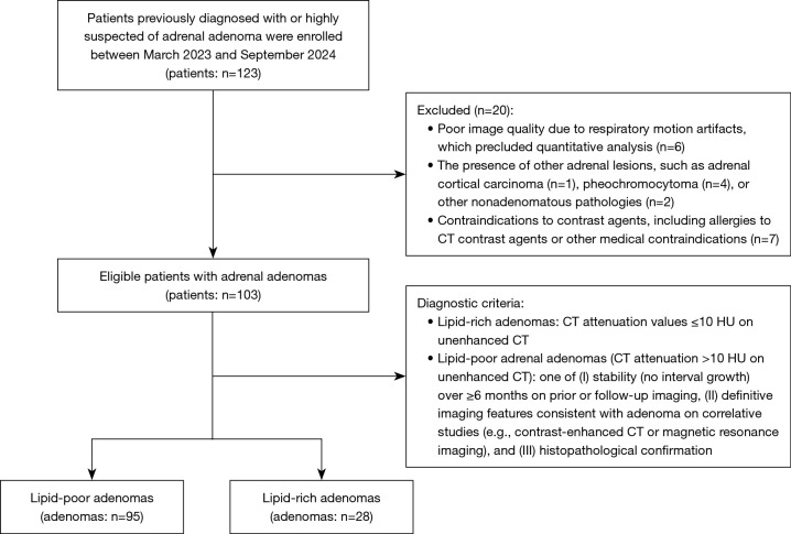
Methods: A total of 103 patients with adrenal adenomas treated at the Adrenal Disease Center of West China Hospital of Sichuan University between March 2023 and September 2024 were enrolled in this prospective study. All patients underwent dual-layer SDCT examination, including TNC and arterial and venous phase scans. VNC images were reconstructed from contrast-enhanced phases. Objective metrics, including CT attenuation value [Hounsfield units (HU)], noise (standard deviation), signal-to-noise ratio (SNR), contrast-to-noise ratio, and absolute attenuation error, and subjective image quality were compared. Interobserver agreement was assessed through the calculation of interclass correlation coefficients. For objective and subjective comparisons between TNC and VNC images, statistical analyses were performed with paired t-tests and Wilcoxon signed-rank tests. The radiation dose with and without TNC was calculated.
Results: This study included 103 patients (48 males and 55 females) with a mean age of 51.33±12.55 years. A total of 123 adrenal adenomas were identified, including 28 lipid-rich adenomas and 95 lipid-poor adenomas. For lipid-poor adenomas, VNC and TNC images showed excellent agreement in CT attenuation values (P>0.05), and compared to VNCp images, VNCa images exhibited significantly lower noise (17.44±3.39 vs. 18.64±2.91 HU; P<0.001) and higher SNR (1.68±0.76 vs. 1.55±0.67; P<0.001). In lipid-rich adenomas, VNC images overestimated CT attenuation, showing high absolute attenuation errors (VNCaerror: 9.92±6.49 HU; VNCperror: 8.50±5.17 HU), although these remained within the acceptable threshold of ≤10 HU. In the subjective scores of image quality, TNC images outperformed VNC images [TNC: median 5, interquartile range (IQR) 5-5; VNC: median 5 (IQR 4-5); P<0.001], although VNC scores remained high. No significant statistical difference was observed between the VNCa and VNCp scores (P>0.05). For most of the surrounding nonadenoma tissues, VNC and TNC images demonstrated good agreement, with attenuation differences consistently within ≤10 HU. Replacing TNC images with VNCa images could reduce the effective dose by approximately 32.63% for lipid-poor adenomas.
Conclusions: Our findings suggest that for lipid-poor adenomas, VNCa demonstrates high agreement with TNC and provides superior image quality, supporting its use as a TNC substitute for reduced radiation dose. For lipid-rich adenomas, VNC should be applied with caution due to the potential risk of attenuation overestimation. Subtype classification remains essential in such studies.

 求助内容:
求助内容: 应助结果提醒方式:


