Nan Wang, Yuhui Liu, Jiangnan Ran, Qi An, Lihua Chen, Ying Zhao, Dan Yu, Ailian Liu, Lina Zhuang, Qingwei Song
{"title":"比较人工智能辅助下呼吸触发T2WI MRI与运动抑制呼吸触发T2WI在腹部成像中的表现。","authors":"Nan Wang, Yuhui Liu, Jiangnan Ran, Qi An, Lihua Chen, Ying Zhao, Dan Yu, Ailian Liu, Lina Zhuang, Qingwei Song","doi":"10.21037/qims-2025-71","DOIUrl":null,"url":null,"abstract":"<p><strong>Background: </strong>Magnetic resonance imaging (MRI) plays a crucial role in the diagnosis of abdominal conditions. A comprehensive assessment, especially of the liver, requires multi-planar T2-weighted sequences. To mitigate the effect of respiratory motion on image quality, the combination of acquisition and reconstruction with motion suppression (ARMS) and respiratory triggering (RT) is commonly employed. While this method maintains image quality, it does so at the expense of longer acquisition times. We evaluated the effectiveness of free-breathing, artificial intelligence-assisted compressed-sensing respiratory-triggered T2-weighted imaging (ACS-RT T2WI) compared to conventional acquisition and reconstruction with motion-suppression respiratory-triggered T2-weighted imaging (ARMS-RT T2WI) in abdominal MRI, assessing both qualitative and quantitative measures of image quality and lesion detection.</p><p><strong>Methods: </strong>In this retrospective study, 334 patients with upper abdominal discomfort were examined on a 3.0T MRI system. Each patient underwent both ARMS-RT T2WI and ACS-RT T2WI. Image quality was analyzed by two independent readers using a five-point Likert scale. The quantitative measurements included the signal-to-noise ratio (SNR), contrast-to-noise ratio (CNR), peak signal-to-noise ratio (PSNR), and sharpness. Lesion detection rates and contrast ratios (CRs) were also evaluated for liver, biliary system, and pancreatic lesions.</p><p><strong>Results: </strong>There ACS-RT T2WI protocol had a significantly reduced median scanning time compared to the ARMS-RT T2WI protocol (148.22±38.37 <i>vs.</i> 13.86±1.72 seconds). However, ARMS-RT T2WI had a higher PSNR than ACS-RT T2WI (39.87±2.72 <i>vs.</i> 38.69±3.00, P<0.05). Of the 201 liver lesions, ARMS-RT T2WI detected 193 (96.0%) and ACS-RT T2WI detected 192 (95.5%) (P=0.787). Of the 97 biliary system lesions, ARMS-RT T2WI detected 92 (94.8%) and ACS-RT T2WI detected 94 (96.9%) (P=0.721). Of the 110 pancreatic lesions, ARMS-RT T2WI detected 102 (92.7%) and ACS-RT T2WI detected 104 (94.5%) (P=0.784). The CR analysis showed the superior performance of ACS-RT T2WI in certain lesion types (hemangioma, 0.58±0.11 <i>vs.</i> 0.55±0.12; biliary tumor, 0.47±0.09 <i>vs.</i> 0.38±0.09; pancreatic cystic lesions, 0.59±0.12 <i>vs.</i> 0.48±0.14; pancreatic cancer, 0.48±0.18 <i>vs.</i> 0.43±0.17), but no significant difference was found in others like focal nodular hyperplasia (FNH), hepatapostema, hepatocellular carcinoma (HCC), cholangiocarcinoma, metastatic tumors, and biliary calculus.</p><p><strong>Conclusions: </strong>ACS-RT T2WI ensures clinical reliability with a substantial scan time reduction (>80%). Despite minor losses in detail and SNR reduction, ACS-RT T2WI does not impair lesion detection, marking its efficacy in abdominal imaging.</p>","PeriodicalId":54267,"journal":{"name":"Quantitative Imaging in Medicine and Surgery","volume":"15 9","pages":"7761-7773"},"PeriodicalIF":2.3000,"publicationDate":"2025-09-01","publicationTypes":"Journal Article","fieldsOfStudy":null,"isOpenAccess":false,"openAccessPdf":"https://www.ncbi.nlm.nih.gov/pmc/articles/PMC12397657/pdf/","citationCount":"0","resultStr":"{\"title\":\"Comparing respiratory-triggered T2WI MRI with an artificial intelligence-assisted technique and motion-suppressed respiratory-triggered T2WI in abdominal imaging.\",\"authors\":\"Nan Wang, Yuhui Liu, Jiangnan Ran, Qi An, Lihua Chen, Ying Zhao, Dan Yu, Ailian Liu, Lina Zhuang, Qingwei Song\",\"doi\":\"10.21037/qims-2025-71\",\"DOIUrl\":null,\"url\":null,\"abstract\":\"<p><strong>Background: </strong>Magnetic resonance imaging (MRI) plays a crucial role in the diagnosis of abdominal conditions. A comprehensive assessment, especially of the liver, requires multi-planar T2-weighted sequences. To mitigate the effect of respiratory motion on image quality, the combination of acquisition and reconstruction with motion suppression (ARMS) and respiratory triggering (RT) is commonly employed. While this method maintains image quality, it does so at the expense of longer acquisition times. We evaluated the effectiveness of free-breathing, artificial intelligence-assisted compressed-sensing respiratory-triggered T2-weighted imaging (ACS-RT T2WI) compared to conventional acquisition and reconstruction with motion-suppression respiratory-triggered T2-weighted imaging (ARMS-RT T2WI) in abdominal MRI, assessing both qualitative and quantitative measures of image quality and lesion detection.</p><p><strong>Methods: </strong>In this retrospective study, 334 patients with upper abdominal discomfort were examined on a 3.0T MRI system. Each patient underwent both ARMS-RT T2WI and ACS-RT T2WI. Image quality was analyzed by two independent readers using a five-point Likert scale. The quantitative measurements included the signal-to-noise ratio (SNR), contrast-to-noise ratio (CNR), peak signal-to-noise ratio (PSNR), and sharpness. Lesion detection rates and contrast ratios (CRs) were also evaluated for liver, biliary system, and pancreatic lesions.</p><p><strong>Results: </strong>There ACS-RT T2WI protocol had a significantly reduced median scanning time compared to the ARMS-RT T2WI protocol (148.22±38.37 <i>vs.</i> 13.86±1.72 seconds). However, ARMS-RT T2WI had a higher PSNR than ACS-RT T2WI (39.87±2.72 <i>vs.</i> 38.69±3.00, P<0.05). Of the 201 liver lesions, ARMS-RT T2WI detected 193 (96.0%) and ACS-RT T2WI detected 192 (95.5%) (P=0.787). Of the 97 biliary system lesions, ARMS-RT T2WI detected 92 (94.8%) and ACS-RT T2WI detected 94 (96.9%) (P=0.721). Of the 110 pancreatic lesions, ARMS-RT T2WI detected 102 (92.7%) and ACS-RT T2WI detected 104 (94.5%) (P=0.784). The CR analysis showed the superior performance of ACS-RT T2WI in certain lesion types (hemangioma, 0.58±0.11 <i>vs.</i> 0.55±0.12; biliary tumor, 0.47±0.09 <i>vs.</i> 0.38±0.09; pancreatic cystic lesions, 0.59±0.12 <i>vs.</i> 0.48±0.14; pancreatic cancer, 0.48±0.18 <i>vs.</i> 0.43±0.17), but no significant difference was found in others like focal nodular hyperplasia (FNH), hepatapostema, hepatocellular carcinoma (HCC), cholangiocarcinoma, metastatic tumors, and biliary calculus.</p><p><strong>Conclusions: </strong>ACS-RT T2WI ensures clinical reliability with a substantial scan time reduction (>80%). Despite minor losses in detail and SNR reduction, ACS-RT T2WI does not impair lesion detection, marking its efficacy in abdominal imaging.</p>\",\"PeriodicalId\":54267,\"journal\":{\"name\":\"Quantitative Imaging in Medicine and Surgery\",\"volume\":\"15 9\",\"pages\":\"7761-7773\"},\"PeriodicalIF\":2.3000,\"publicationDate\":\"2025-09-01\",\"publicationTypes\":\"Journal Article\",\"fieldsOfStudy\":null,\"isOpenAccess\":false,\"openAccessPdf\":\"https://www.ncbi.nlm.nih.gov/pmc/articles/PMC12397657/pdf/\",\"citationCount\":\"0\",\"resultStr\":null,\"platform\":\"Semanticscholar\",\"paperid\":null,\"PeriodicalName\":\"Quantitative Imaging in Medicine and Surgery\",\"FirstCategoryId\":\"3\",\"ListUrlMain\":\"https://doi.org/10.21037/qims-2025-71\",\"RegionNum\":2,\"RegionCategory\":\"医学\",\"ArticlePicture\":[],\"TitleCN\":null,\"AbstractTextCN\":null,\"PMCID\":null,\"EPubDate\":\"2025/8/19 0:00:00\",\"PubModel\":\"Epub\",\"JCR\":\"Q2\",\"JCRName\":\"RADIOLOGY, NUCLEAR MEDICINE & MEDICAL IMAGING\",\"Score\":null,\"Total\":0}","platform":"Semanticscholar","paperid":null,"PeriodicalName":"Quantitative Imaging in Medicine and Surgery","FirstCategoryId":"3","ListUrlMain":"https://doi.org/10.21037/qims-2025-71","RegionNum":2,"RegionCategory":"医学","ArticlePicture":[],"TitleCN":null,"AbstractTextCN":null,"PMCID":null,"EPubDate":"2025/8/19 0:00:00","PubModel":"Epub","JCR":"Q2","JCRName":"RADIOLOGY, NUCLEAR MEDICINE & MEDICAL IMAGING","Score":null,"Total":0}
引用次数: 0
摘要
背景:磁共振成像(MRI)在腹部疾病的诊断中起着至关重要的作用。全面评估,尤其是肝脏,需要多平面t2加权序列。为了减轻呼吸运动对图像质量的影响,通常采用运动抑制(ARMS)和呼吸触发(RT)相结合的采集和重建方法。虽然这种方法可以保持图像质量,但这样做的代价是更长的采集时间。我们评估了自由呼吸,人工智能辅助压缩传感呼吸触发t2加权成像(ACS-RT T2WI)与常规采集和重建运动抑制呼吸触发t2加权成像(ARMS-RT T2WI)在腹部MRI中的有效性,评估了图像质量和病变检测的定性和定量指标。方法:对334例上腹部不适患者在3.0T MRI系统上进行回顾性研究。每位患者均接受ARMS-RT T2WI和ACS-RT T2WI检查。图像质量由两个独立的读者使用五点李克特量表进行分析。定量测量包括信噪比(SNR)、噪声对比比(CNR)、峰值信噪比(PSNR)和清晰度。同时评估肝脏、胆道系统和胰腺病变的检出率和对比比(CRs)。结果:ACS-RT T2WI方案的中位扫描时间较ARMS-RT T2WI方案显著缩短(148.22±38.37秒vs 13.86±1.72秒)。然而,ARMS-RT T2WI的PSNR高于ACS-RT T2WI(39.87±2.72 vs. 38.69±3.00,PSNR为0.55±0.12;胆道肿瘤为0.47±0.09 vs. 0.38±0.09;胰腺囊性病变为0.59±0.12 vs. 0.48±0.14;胰腺癌为0.48±0.18 vs. 0.43±0.17),但在局灶性结节性增生(FNH)、肝水肿、肝细胞癌(HCC)、胆管癌、转移性肿瘤、胆道结石等方面无显著差异。结论:ACS-RT T2WI可大幅缩短扫描时间(bbb80 %),确保临床可靠性。尽管有细微的细节损失和信噪比降低,但ACS-RT T2WI并不影响病变的检测,表明其在腹部成像中的有效性。
Comparing respiratory-triggered T2WI MRI with an artificial intelligence-assisted technique and motion-suppressed respiratory-triggered T2WI in abdominal imaging.
Background: Magnetic resonance imaging (MRI) plays a crucial role in the diagnosis of abdominal conditions. A comprehensive assessment, especially of the liver, requires multi-planar T2-weighted sequences. To mitigate the effect of respiratory motion on image quality, the combination of acquisition and reconstruction with motion suppression (ARMS) and respiratory triggering (RT) is commonly employed. While this method maintains image quality, it does so at the expense of longer acquisition times. We evaluated the effectiveness of free-breathing, artificial intelligence-assisted compressed-sensing respiratory-triggered T2-weighted imaging (ACS-RT T2WI) compared to conventional acquisition and reconstruction with motion-suppression respiratory-triggered T2-weighted imaging (ARMS-RT T2WI) in abdominal MRI, assessing both qualitative and quantitative measures of image quality and lesion detection.
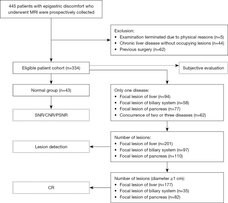
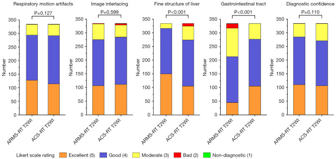
Methods: In this retrospective study, 334 patients with upper abdominal discomfort were examined on a 3.0T MRI system. Each patient underwent both ARMS-RT T2WI and ACS-RT T2WI. Image quality was analyzed by two independent readers using a five-point Likert scale. The quantitative measurements included the signal-to-noise ratio (SNR), contrast-to-noise ratio (CNR), peak signal-to-noise ratio (PSNR), and sharpness. Lesion detection rates and contrast ratios (CRs) were also evaluated for liver, biliary system, and pancreatic lesions.
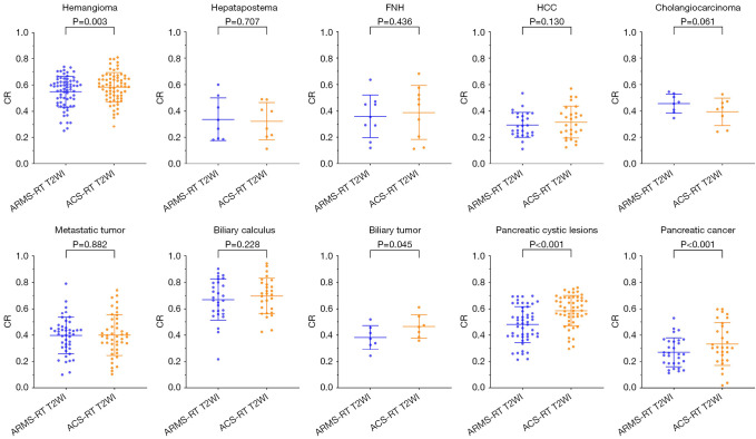
Results: There ACS-RT T2WI protocol had a significantly reduced median scanning time compared to the ARMS-RT T2WI protocol (148.22±38.37 vs. 13.86±1.72 seconds). However, ARMS-RT T2WI had a higher PSNR than ACS-RT T2WI (39.87±2.72 vs. 38.69±3.00, P<0.05). Of the 201 liver lesions, ARMS-RT T2WI detected 193 (96.0%) and ACS-RT T2WI detected 192 (95.5%) (P=0.787). Of the 97 biliary system lesions, ARMS-RT T2WI detected 92 (94.8%) and ACS-RT T2WI detected 94 (96.9%) (P=0.721). Of the 110 pancreatic lesions, ARMS-RT T2WI detected 102 (92.7%) and ACS-RT T2WI detected 104 (94.5%) (P=0.784). The CR analysis showed the superior performance of ACS-RT T2WI in certain lesion types (hemangioma, 0.58±0.11 vs. 0.55±0.12; biliary tumor, 0.47±0.09 vs. 0.38±0.09; pancreatic cystic lesions, 0.59±0.12 vs. 0.48±0.14; pancreatic cancer, 0.48±0.18 vs. 0.43±0.17), but no significant difference was found in others like focal nodular hyperplasia (FNH), hepatapostema, hepatocellular carcinoma (HCC), cholangiocarcinoma, metastatic tumors, and biliary calculus.
Conclusions: ACS-RT T2WI ensures clinical reliability with a substantial scan time reduction (>80%). Despite minor losses in detail and SNR reduction, ACS-RT T2WI does not impair lesion detection, marking its efficacy in abdominal imaging.

 求助内容:
求助内容: 应助结果提醒方式:


