Peiming Qin, Yan Yi, Cheng Xu, Limiao Zou, Fenggang Jia, Jian Guo, Ming Wang, Yan Zhang, Ziquan Wang, Pei Dong, Dijia Wu, Xiaodong Wang, Yining Wang
{"title":"利用心肌ct灌注信息优化分流储备计算。","authors":"Peiming Qin, Yan Yi, Cheng Xu, Limiao Zou, Fenggang Jia, Jian Guo, Ming Wang, Yan Zhang, Ziquan Wang, Pei Dong, Dijia Wu, Xiaodong Wang, Yining Wang","doi":"10.21037/qims-24-2172","DOIUrl":null,"url":null,"abstract":"<p><strong>Background: </strong>Current computed tomography angiography-derived fractional flow reserve (CT-FFR) diagnosis leaves room for improvement in diagnosing coronary heart disease. In this study, the computed fluid dynamics boundary condition optimization method was used to calculate CT-FFR aiming to improve the diagnostic accuracy of CT-FFR for coronary heart disease.</p><p><strong>Methods: </strong>The two enhancement approaches are as follows: (A) inlet flow optimization, which involves determining the total coronary inlet flow rate by summing the myocardial blood flow (MBF) across the entire left ventricular myocardium; and (B) inlet & outlet flow optimization: building upon method A, where the outlet flow of coronary artery branches is calculated through blood supply area analysis.</p><p><strong>Results: </strong>A total of 100 fractional flow reserve pressure guide wire measurement sites from 47 cases were used to evaluate the above two methods comparing with the traditional computed fluid dynamics method without computed tomography perfusion (CTP) images. In traditional method, the accuracy was 88%, the sensitivity was 91.4% (95% confidence interval: 75.8-97.7%), and the specificity was 86.2% (95% confidence interval: 74.8-93.1%). In Method A, the accuracy improved by 5% (93%), the sensitivity remained unchanged (91.4%, 95% confidence interval: 75.8-97.7%), and the specificity increased by 7.6% (93.8%, 95% confidence interval: 84.2-98%). In Method B, the accuracy increased by 6% (94%), the sensitivity increased to 100% (95% confidence interval: 87.7-100%), and the specificity increased by 4.8% (94%, 95% confidence interval: 80.3-96.2%).</p><p><strong>Conclusions: </strong>The computed fluid dynamics calculation, guided by MBF values from stress CTP imaging, helps enhance the consistency between CT-FFR calculation and invasive fractional flow reserve measurements.</p>","PeriodicalId":54267,"journal":{"name":"Quantitative Imaging in Medicine and Surgery","volume":"15 9","pages":"7909-7921"},"PeriodicalIF":2.3000,"publicationDate":"2025-09-01","publicationTypes":"Journal Article","fieldsOfStudy":null,"isOpenAccess":false,"openAccessPdf":"https://www.ncbi.nlm.nih.gov/pmc/articles/PMC12397671/pdf/","citationCount":"0","resultStr":"{\"title\":\"Fractional flow reserve calculation optimized by myocardial computed tomography perfusion information.\",\"authors\":\"Peiming Qin, Yan Yi, Cheng Xu, Limiao Zou, Fenggang Jia, Jian Guo, Ming Wang, Yan Zhang, Ziquan Wang, Pei Dong, Dijia Wu, Xiaodong Wang, Yining Wang\",\"doi\":\"10.21037/qims-24-2172\",\"DOIUrl\":null,\"url\":null,\"abstract\":\"<p><strong>Background: </strong>Current computed tomography angiography-derived fractional flow reserve (CT-FFR) diagnosis leaves room for improvement in diagnosing coronary heart disease. In this study, the computed fluid dynamics boundary condition optimization method was used to calculate CT-FFR aiming to improve the diagnostic accuracy of CT-FFR for coronary heart disease.</p><p><strong>Methods: </strong>The two enhancement approaches are as follows: (A) inlet flow optimization, which involves determining the total coronary inlet flow rate by summing the myocardial blood flow (MBF) across the entire left ventricular myocardium; and (B) inlet & outlet flow optimization: building upon method A, where the outlet flow of coronary artery branches is calculated through blood supply area analysis.</p><p><strong>Results: </strong>A total of 100 fractional flow reserve pressure guide wire measurement sites from 47 cases were used to evaluate the above two methods comparing with the traditional computed fluid dynamics method without computed tomography perfusion (CTP) images. In traditional method, the accuracy was 88%, the sensitivity was 91.4% (95% confidence interval: 75.8-97.7%), and the specificity was 86.2% (95% confidence interval: 74.8-93.1%). In Method A, the accuracy improved by 5% (93%), the sensitivity remained unchanged (91.4%, 95% confidence interval: 75.8-97.7%), and the specificity increased by 7.6% (93.8%, 95% confidence interval: 84.2-98%). In Method B, the accuracy increased by 6% (94%), the sensitivity increased to 100% (95% confidence interval: 87.7-100%), and the specificity increased by 4.8% (94%, 95% confidence interval: 80.3-96.2%).</p><p><strong>Conclusions: </strong>The computed fluid dynamics calculation, guided by MBF values from stress CTP imaging, helps enhance the consistency between CT-FFR calculation and invasive fractional flow reserve measurements.</p>\",\"PeriodicalId\":54267,\"journal\":{\"name\":\"Quantitative Imaging in Medicine and Surgery\",\"volume\":\"15 9\",\"pages\":\"7909-7921\"},\"PeriodicalIF\":2.3000,\"publicationDate\":\"2025-09-01\",\"publicationTypes\":\"Journal Article\",\"fieldsOfStudy\":null,\"isOpenAccess\":false,\"openAccessPdf\":\"https://www.ncbi.nlm.nih.gov/pmc/articles/PMC12397671/pdf/\",\"citationCount\":\"0\",\"resultStr\":null,\"platform\":\"Semanticscholar\",\"paperid\":null,\"PeriodicalName\":\"Quantitative Imaging in Medicine and Surgery\",\"FirstCategoryId\":\"3\",\"ListUrlMain\":\"https://doi.org/10.21037/qims-24-2172\",\"RegionNum\":2,\"RegionCategory\":\"医学\",\"ArticlePicture\":[],\"TitleCN\":null,\"AbstractTextCN\":null,\"PMCID\":null,\"EPubDate\":\"2025/8/19 0:00:00\",\"PubModel\":\"Epub\",\"JCR\":\"Q2\",\"JCRName\":\"RADIOLOGY, NUCLEAR MEDICINE & MEDICAL IMAGING\",\"Score\":null,\"Total\":0}","platform":"Semanticscholar","paperid":null,"PeriodicalName":"Quantitative Imaging in Medicine and Surgery","FirstCategoryId":"3","ListUrlMain":"https://doi.org/10.21037/qims-24-2172","RegionNum":2,"RegionCategory":"医学","ArticlePicture":[],"TitleCN":null,"AbstractTextCN":null,"PMCID":null,"EPubDate":"2025/8/19 0:00:00","PubModel":"Epub","JCR":"Q2","JCRName":"RADIOLOGY, NUCLEAR MEDICINE & MEDICAL IMAGING","Score":null,"Total":0}
Background: Current computed tomography angiography-derived fractional flow reserve (CT-FFR) diagnosis leaves room for improvement in diagnosing coronary heart disease. In this study, the computed fluid dynamics boundary condition optimization method was used to calculate CT-FFR aiming to improve the diagnostic accuracy of CT-FFR for coronary heart disease.
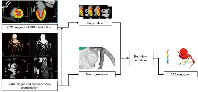
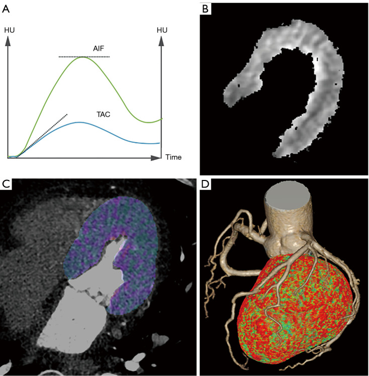
Methods: The two enhancement approaches are as follows: (A) inlet flow optimization, which involves determining the total coronary inlet flow rate by summing the myocardial blood flow (MBF) across the entire left ventricular myocardium; and (B) inlet & outlet flow optimization: building upon method A, where the outlet flow of coronary artery branches is calculated through blood supply area analysis.
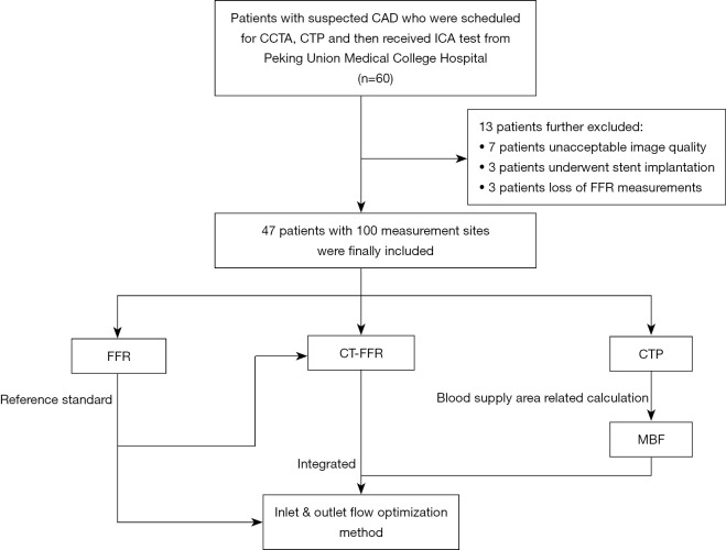
Results: A total of 100 fractional flow reserve pressure guide wire measurement sites from 47 cases were used to evaluate the above two methods comparing with the traditional computed fluid dynamics method without computed tomography perfusion (CTP) images. In traditional method, the accuracy was 88%, the sensitivity was 91.4% (95% confidence interval: 75.8-97.7%), and the specificity was 86.2% (95% confidence interval: 74.8-93.1%). In Method A, the accuracy improved by 5% (93%), the sensitivity remained unchanged (91.4%, 95% confidence interval: 75.8-97.7%), and the specificity increased by 7.6% (93.8%, 95% confidence interval: 84.2-98%). In Method B, the accuracy increased by 6% (94%), the sensitivity increased to 100% (95% confidence interval: 87.7-100%), and the specificity increased by 4.8% (94%, 95% confidence interval: 80.3-96.2%).
Conclusions: The computed fluid dynamics calculation, guided by MBF values from stress CTP imaging, helps enhance the consistency between CT-FFR calculation and invasive fractional flow reserve measurements.

 求助内容:
求助内容: 应助结果提醒方式:


