Xinyan Wu, Ling Yang, Li Yu, Lingqin Zhang, Nian Liu, Xiaojun Lu, Kang Li
{"title":"克罗恩病和腹痛患者的异常结构改变和功能连接紊乱:基于体素的形态测量和功能磁共振成像研究","authors":"Xinyan Wu, Ling Yang, Li Yu, Lingqin Zhang, Nian Liu, Xiaojun Lu, Kang Li","doi":"10.21037/qims-2024-2572","DOIUrl":null,"url":null,"abstract":"<p><strong>Background: </strong>Abdominal pain is a prevalent and debilitating manifestation of Crohn's disease (CD) that significantly impacts the lives of those affected. The neurological pathways responsible for abdominal pain in patients with CD remain unidentified. Therefore, the purpose of this study was to characterize the structural alterations in the brain and associated functional connectivity (FC) in patients with CD and abdominal pain.</p><p><strong>Methods: </strong>The data for three-dimensional T1-weighted and resting-state functional magnetic resonance imaging (fMRI) were gathered from 23 patients with CD and abdominal pain (pain CD), 24 patients with CD but without abdominal pain (nonpain CD), and 25 healthy controls (HCs). Differences in gray-matter volume (GMV) and FC between the pain CD group, nonpain CD group, and HCs were evaluated via analysis of covariance. Biased correlation analyses were employed to evaluate the association of variations in GMV and FC with clinical measures.</p><p><strong>Results: </strong>Voxel-based morphometry analysis revealed that the pain CD group exhibited changes in GMV in the right anterior cingulate cortex (ACC) and orbitofrontal regions, including the orbital parts of the superior frontal gyri, middle frontal gyri (ORBmid), and inferior frontal gyri, as compared to both the HC and nonpain CD groups. Additionally, compared to the HC group, the nonpain CD group showed increased GMV in the bilateral hippocampus. FC analysis showed that the pain CD group had enhanced FC between the right ACC and the default mode network (DMN), particularly with the parahippocampal gyrus (PHG), Rolandic operculum, and postcentral gyrus, as compared to the nonpain CD group. Furthermore, compared to both the nonpain CD and HC groups, pain CD group exhibited increased FC between the left ORBmid and key pain-processing hubs, including the left thalamus, left ACC, and right middle frontal gyrus (MFG). Notably, the FC between the ACC and PHG was negatively correlated with Beck Depression Inventory score (r=-0.548; P=0.019). The FC between the left ORBmid and the right MFG showed a significant negative correlation with Pain Sensitivity Questionnaire score (r=-0.495; P=0.037).</p><p><strong>Conclusions: </strong>Our results suggest that pain may differentially affect brain morphology and function in patients with CD, particularly involving the ACC and orbitofrontal cortex. Specifically, increased FC between the ACC and DMN, as well as orbitofrontal-thalamic circuits, provide novel imaging evidence for the neural mechanisms underlying visceral pain in CD.</p>","PeriodicalId":54267,"journal":{"name":"Quantitative Imaging in Medicine and Surgery","volume":"15 9","pages":"8265-8281"},"PeriodicalIF":2.3000,"publicationDate":"2025-09-01","publicationTypes":"Journal Article","fieldsOfStudy":null,"isOpenAccess":false,"openAccessPdf":"https://www.ncbi.nlm.nih.gov/pmc/articles/PMC12397677/pdf/","citationCount":"0","resultStr":"{\"title\":\"Abnormal structural changes and disturbed functional connectivity in patients with Crohn's disease and abdominal pain: a voxel-based morphometry and functional magnetic resonance imaging study.\",\"authors\":\"Xinyan Wu, Ling Yang, Li Yu, Lingqin Zhang, Nian Liu, Xiaojun Lu, Kang Li\",\"doi\":\"10.21037/qims-2024-2572\",\"DOIUrl\":null,\"url\":null,\"abstract\":\"<p><strong>Background: </strong>Abdominal pain is a prevalent and debilitating manifestation of Crohn's disease (CD) that significantly impacts the lives of those affected. The neurological pathways responsible for abdominal pain in patients with CD remain unidentified. Therefore, the purpose of this study was to characterize the structural alterations in the brain and associated functional connectivity (FC) in patients with CD and abdominal pain.</p><p><strong>Methods: </strong>The data for three-dimensional T1-weighted and resting-state functional magnetic resonance imaging (fMRI) were gathered from 23 patients with CD and abdominal pain (pain CD), 24 patients with CD but without abdominal pain (nonpain CD), and 25 healthy controls (HCs). Differences in gray-matter volume (GMV) and FC between the pain CD group, nonpain CD group, and HCs were evaluated via analysis of covariance. Biased correlation analyses were employed to evaluate the association of variations in GMV and FC with clinical measures.</p><p><strong>Results: </strong>Voxel-based morphometry analysis revealed that the pain CD group exhibited changes in GMV in the right anterior cingulate cortex (ACC) and orbitofrontal regions, including the orbital parts of the superior frontal gyri, middle frontal gyri (ORBmid), and inferior frontal gyri, as compared to both the HC and nonpain CD groups. Additionally, compared to the HC group, the nonpain CD group showed increased GMV in the bilateral hippocampus. FC analysis showed that the pain CD group had enhanced FC between the right ACC and the default mode network (DMN), particularly with the parahippocampal gyrus (PHG), Rolandic operculum, and postcentral gyrus, as compared to the nonpain CD group. Furthermore, compared to both the nonpain CD and HC groups, pain CD group exhibited increased FC between the left ORBmid and key pain-processing hubs, including the left thalamus, left ACC, and right middle frontal gyrus (MFG). Notably, the FC between the ACC and PHG was negatively correlated with Beck Depression Inventory score (r=-0.548; P=0.019). The FC between the left ORBmid and the right MFG showed a significant negative correlation with Pain Sensitivity Questionnaire score (r=-0.495; P=0.037).</p><p><strong>Conclusions: </strong>Our results suggest that pain may differentially affect brain morphology and function in patients with CD, particularly involving the ACC and orbitofrontal cortex. Specifically, increased FC between the ACC and DMN, as well as orbitofrontal-thalamic circuits, provide novel imaging evidence for the neural mechanisms underlying visceral pain in CD.</p>\",\"PeriodicalId\":54267,\"journal\":{\"name\":\"Quantitative Imaging in Medicine and Surgery\",\"volume\":\"15 9\",\"pages\":\"8265-8281\"},\"PeriodicalIF\":2.3000,\"publicationDate\":\"2025-09-01\",\"publicationTypes\":\"Journal Article\",\"fieldsOfStudy\":null,\"isOpenAccess\":false,\"openAccessPdf\":\"https://www.ncbi.nlm.nih.gov/pmc/articles/PMC12397677/pdf/\",\"citationCount\":\"0\",\"resultStr\":null,\"platform\":\"Semanticscholar\",\"paperid\":null,\"PeriodicalName\":\"Quantitative Imaging in Medicine and Surgery\",\"FirstCategoryId\":\"3\",\"ListUrlMain\":\"https://doi.org/10.21037/qims-2024-2572\",\"RegionNum\":2,\"RegionCategory\":\"医学\",\"ArticlePicture\":[],\"TitleCN\":null,\"AbstractTextCN\":null,\"PMCID\":null,\"EPubDate\":\"2025/8/13 0:00:00\",\"PubModel\":\"Epub\",\"JCR\":\"Q2\",\"JCRName\":\"RADIOLOGY, NUCLEAR MEDICINE & MEDICAL IMAGING\",\"Score\":null,\"Total\":0}","platform":"Semanticscholar","paperid":null,"PeriodicalName":"Quantitative Imaging in Medicine and Surgery","FirstCategoryId":"3","ListUrlMain":"https://doi.org/10.21037/qims-2024-2572","RegionNum":2,"RegionCategory":"医学","ArticlePicture":[],"TitleCN":null,"AbstractTextCN":null,"PMCID":null,"EPubDate":"2025/8/13 0:00:00","PubModel":"Epub","JCR":"Q2","JCRName":"RADIOLOGY, NUCLEAR MEDICINE & MEDICAL IMAGING","Score":null,"Total":0}
Abnormal structural changes and disturbed functional connectivity in patients with Crohn's disease and abdominal pain: a voxel-based morphometry and functional magnetic resonance imaging study.
Background: Abdominal pain is a prevalent and debilitating manifestation of Crohn's disease (CD) that significantly impacts the lives of those affected. The neurological pathways responsible for abdominal pain in patients with CD remain unidentified. Therefore, the purpose of this study was to characterize the structural alterations in the brain and associated functional connectivity (FC) in patients with CD and abdominal pain.
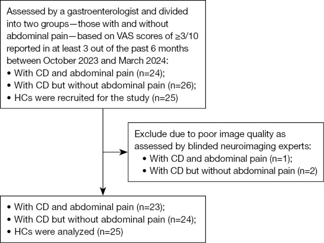
Methods: The data for three-dimensional T1-weighted and resting-state functional magnetic resonance imaging (fMRI) were gathered from 23 patients with CD and abdominal pain (pain CD), 24 patients with CD but without abdominal pain (nonpain CD), and 25 healthy controls (HCs). Differences in gray-matter volume (GMV) and FC between the pain CD group, nonpain CD group, and HCs were evaluated via analysis of covariance. Biased correlation analyses were employed to evaluate the association of variations in GMV and FC with clinical measures.
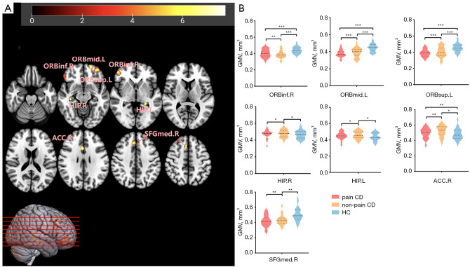
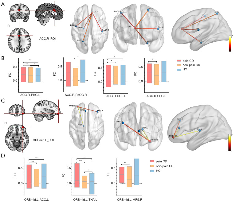
Results: Voxel-based morphometry analysis revealed that the pain CD group exhibited changes in GMV in the right anterior cingulate cortex (ACC) and orbitofrontal regions, including the orbital parts of the superior frontal gyri, middle frontal gyri (ORBmid), and inferior frontal gyri, as compared to both the HC and nonpain CD groups. Additionally, compared to the HC group, the nonpain CD group showed increased GMV in the bilateral hippocampus. FC analysis showed that the pain CD group had enhanced FC between the right ACC and the default mode network (DMN), particularly with the parahippocampal gyrus (PHG), Rolandic operculum, and postcentral gyrus, as compared to the nonpain CD group. Furthermore, compared to both the nonpain CD and HC groups, pain CD group exhibited increased FC between the left ORBmid and key pain-processing hubs, including the left thalamus, left ACC, and right middle frontal gyrus (MFG). Notably, the FC between the ACC and PHG was negatively correlated with Beck Depression Inventory score (r=-0.548; P=0.019). The FC between the left ORBmid and the right MFG showed a significant negative correlation with Pain Sensitivity Questionnaire score (r=-0.495; P=0.037).
Conclusions: Our results suggest that pain may differentially affect brain morphology and function in patients with CD, particularly involving the ACC and orbitofrontal cortex. Specifically, increased FC between the ACC and DMN, as well as orbitofrontal-thalamic circuits, provide novel imaging evidence for the neural mechanisms underlying visceral pain in CD.

 求助内容:
求助内容: 应助结果提醒方式:


