Elaheh Mianehsaz, Hamidreza Talari, Marziyeh Naghavi Ravandi, Mohammad Hossein Tabatabaei, Mohammad Javad Azadchehr, Saeedeh Eshraqi, Mohammad Mahdi Heidari
{"title":"评估各种超声标准确定腕管综合征的严重程度。","authors":"Elaheh Mianehsaz, Hamidreza Talari, Marziyeh Naghavi Ravandi, Mohammad Hossein Tabatabaei, Mohammad Javad Azadchehr, Saeedeh Eshraqi, Mohammad Mahdi Heidari","doi":"10.1155/rrp/4936187","DOIUrl":null,"url":null,"abstract":"<p><p><b>Objective:</b> This study aimed at assessing the value of a variety of ultrasound criteria for grading carpal tunnel syndrome (CTS) severity. <b>Methods:</b> Ultrasound evaluations were conducted on confirmed CTS patients by an experienced radiologist, blinded to NCS results. Cross-sectional area (CSA) at pronator quadratus muscle, carpal tunnel inlet and outlet, echogenicity, transverse motion during flexion, flattening ratio, and thickening of the flexor retinaculum were measured. <b>Results:</b> Decreased echogenicity of the median nerve was notably observed as a distinguishing feature between mild and moderate cases. Decreased nerve movement was significantly more prevalent in severe CTS cases. No significant differences were found in the median nerve flattening ratio or flexor retinaculum thickness. Bowing at the inlet showed significant differences. CSA at the inlet and outlet indicated severe CTS with significant differences. <b>Conclusion:</b> The findings highlight the importance of using multiple sonographic criteria for accurate diagnosis and treatment, although no significant differences were noted in the median nerve flattening ratio and flexor retinaculum thickness.</p>","PeriodicalId":51864,"journal":{"name":"Radiology Research and Practice","volume":"2025 ","pages":"4936187"},"PeriodicalIF":1.5000,"publicationDate":"2025-08-19","publicationTypes":"Journal Article","fieldsOfStudy":null,"isOpenAccess":false,"openAccessPdf":"https://www.ncbi.nlm.nih.gov/pmc/articles/PMC12380511/pdf/","citationCount":"0","resultStr":"{\"title\":\"Evaluating Various Ultrasound Criteria for Determining Carpal Tunnel Syndrome Severity.\",\"authors\":\"Elaheh Mianehsaz, Hamidreza Talari, Marziyeh Naghavi Ravandi, Mohammad Hossein Tabatabaei, Mohammad Javad Azadchehr, Saeedeh Eshraqi, Mohammad Mahdi Heidari\",\"doi\":\"10.1155/rrp/4936187\",\"DOIUrl\":null,\"url\":null,\"abstract\":\"<p><p><b>Objective:</b> This study aimed at assessing the value of a variety of ultrasound criteria for grading carpal tunnel syndrome (CTS) severity. <b>Methods:</b> Ultrasound evaluations were conducted on confirmed CTS patients by an experienced radiologist, blinded to NCS results. Cross-sectional area (CSA) at pronator quadratus muscle, carpal tunnel inlet and outlet, echogenicity, transverse motion during flexion, flattening ratio, and thickening of the flexor retinaculum were measured. <b>Results:</b> Decreased echogenicity of the median nerve was notably observed as a distinguishing feature between mild and moderate cases. Decreased nerve movement was significantly more prevalent in severe CTS cases. No significant differences were found in the median nerve flattening ratio or flexor retinaculum thickness. Bowing at the inlet showed significant differences. CSA at the inlet and outlet indicated severe CTS with significant differences. <b>Conclusion:</b> The findings highlight the importance of using multiple sonographic criteria for accurate diagnosis and treatment, although no significant differences were noted in the median nerve flattening ratio and flexor retinaculum thickness.</p>\",\"PeriodicalId\":51864,\"journal\":{\"name\":\"Radiology Research and Practice\",\"volume\":\"2025 \",\"pages\":\"4936187\"},\"PeriodicalIF\":1.5000,\"publicationDate\":\"2025-08-19\",\"publicationTypes\":\"Journal Article\",\"fieldsOfStudy\":null,\"isOpenAccess\":false,\"openAccessPdf\":\"https://www.ncbi.nlm.nih.gov/pmc/articles/PMC12380511/pdf/\",\"citationCount\":\"0\",\"resultStr\":null,\"platform\":\"Semanticscholar\",\"paperid\":null,\"PeriodicalName\":\"Radiology Research and Practice\",\"FirstCategoryId\":\"1085\",\"ListUrlMain\":\"https://doi.org/10.1155/rrp/4936187\",\"RegionNum\":0,\"RegionCategory\":null,\"ArticlePicture\":[],\"TitleCN\":null,\"AbstractTextCN\":null,\"PMCID\":null,\"EPubDate\":\"2025/1/1 0:00:00\",\"PubModel\":\"eCollection\",\"JCR\":\"Q2\",\"JCRName\":\"RADIOLOGY, NUCLEAR MEDICINE & MEDICAL IMAGING\",\"Score\":null,\"Total\":0}","platform":"Semanticscholar","paperid":null,"PeriodicalName":"Radiology Research and Practice","FirstCategoryId":"1085","ListUrlMain":"https://doi.org/10.1155/rrp/4936187","RegionNum":0,"RegionCategory":null,"ArticlePicture":[],"TitleCN":null,"AbstractTextCN":null,"PMCID":null,"EPubDate":"2025/1/1 0:00:00","PubModel":"eCollection","JCR":"Q2","JCRName":"RADIOLOGY, NUCLEAR MEDICINE & MEDICAL IMAGING","Score":null,"Total":0}
Evaluating Various Ultrasound Criteria for Determining Carpal Tunnel Syndrome Severity.
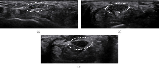
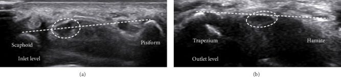
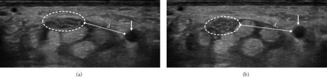
Objective: This study aimed at assessing the value of a variety of ultrasound criteria for grading carpal tunnel syndrome (CTS) severity. Methods: Ultrasound evaluations were conducted on confirmed CTS patients by an experienced radiologist, blinded to NCS results. Cross-sectional area (CSA) at pronator quadratus muscle, carpal tunnel inlet and outlet, echogenicity, transverse motion during flexion, flattening ratio, and thickening of the flexor retinaculum were measured. Results: Decreased echogenicity of the median nerve was notably observed as a distinguishing feature between mild and moderate cases. Decreased nerve movement was significantly more prevalent in severe CTS cases. No significant differences were found in the median nerve flattening ratio or flexor retinaculum thickness. Bowing at the inlet showed significant differences. CSA at the inlet and outlet indicated severe CTS with significant differences. Conclusion: The findings highlight the importance of using multiple sonographic criteria for accurate diagnosis and treatment, although no significant differences were noted in the median nerve flattening ratio and flexor retinaculum thickness.
期刊介绍:
Radiology Research and Practice is a peer-reviewed, Open Access journal that publishes articles on all areas of medical imaging. The journal promotes evidence-based radiology practice though the publication of original research, reviews, and clinical studies for a multidisciplinary audience. Radiology Research and Practice is archived in Portico, which provides permanent archiving for electronic scholarly journals, as well as via the LOCKSS initiative. It operates a fully open access publishing model which allows open global access to its published content. This model is supported through Article Processing Charges. For more information on Article Processing charges in gen

 求助内容:
求助内容: 应助结果提醒方式:


