John Kendall, Gabriel Gaspar, Derek Berger, Jacob Levman
{"title":"小儿阑尾炎的机器学习与特征选择。","authors":"John Kendall, Gabriel Gaspar, Derek Berger, Jacob Levman","doi":"10.3390/tomography11080090","DOIUrl":null,"url":null,"abstract":"<p><strong>Background/objectives: </strong>Accurate prediction of pediatric appendicitis diagnosis, management, and severity is critical for clinical decision-making. We aimed to evaluate the predictive performance of a wide range of machine learning models, combined with various feature selection techniques, on a pediatric appendicitis dataset. A particular focus was placed on the role of ultrasound (US) image-descriptive features in model performance and explainability.</p><p><strong>Methods: </strong>We conducted a retrospective cohort study on a dataset of 781 pediatric patients aged 0-18 presenting to Children's Hospital St. Hedwig in Regensburg, Germany, between January 2016 and February 2023. We developed and validated predictive models; machine learning algorithms included the random forest, logistic regression, stochastic gradient descent, and the light gradient boosting machine (LGBM). These were paired exhaustively with feature selection methods spanning filter-based (association and prediction), embedded (LGBM and linear), and a novel redundancy-aware step-up wrapper approach. We employed a machine learning benchmarking study design where AI models were trained to predict diagnosis, management, and severity outcomes, both with and without US image-descriptive features, and evaluated on held-out testing samples. Model performance was assessed using overall accuracy and area under the receiver operating characteristic curve (AUROC). A deep learner optimized for tabular data, GANDALF, was also evaluated in these applications.</p><p><strong>Results: </strong>US features significantly improved diagnostic accuracy, supporting their use in reducing model bias. However, they were not essential for maximizing accuracy in predicting management or severity. In summary, our best-performing models were, for diagnosis, the random forest with embedded LGBM feature selection (98.1% accuracy, AUROC: 0.993), for management, the random forest without feature selection (93.9% accuracy, AUROC: 0.980), and for severity, the LGBM with filter-based association feature selection (90.1% accuracy, AUROC: 0.931).</p><p><strong>Conclusions: </strong>Our results demonstrate that high-performing, interpretable machine learning models can predict key clinical outcomes in pediatric appendicitis. US image features improve diagnostic accuracy but are not critical for predicting management or severity.</p>","PeriodicalId":51330,"journal":{"name":"Tomography","volume":"11 8","pages":""},"PeriodicalIF":2.2000,"publicationDate":"2025-08-13","publicationTypes":"Journal Article","fieldsOfStudy":null,"isOpenAccess":false,"openAccessPdf":"https://www.ncbi.nlm.nih.gov/pmc/articles/PMC12390108/pdf/","citationCount":"0","resultStr":"{\"title\":\"Machine Learning and Feature Selection in Pediatric Appendicitis.\",\"authors\":\"John Kendall, Gabriel Gaspar, Derek Berger, Jacob Levman\",\"doi\":\"10.3390/tomography11080090\",\"DOIUrl\":null,\"url\":null,\"abstract\":\"<p><strong>Background/objectives: </strong>Accurate prediction of pediatric appendicitis diagnosis, management, and severity is critical for clinical decision-making. We aimed to evaluate the predictive performance of a wide range of machine learning models, combined with various feature selection techniques, on a pediatric appendicitis dataset. A particular focus was placed on the role of ultrasound (US) image-descriptive features in model performance and explainability.</p><p><strong>Methods: </strong>We conducted a retrospective cohort study on a dataset of 781 pediatric patients aged 0-18 presenting to Children's Hospital St. Hedwig in Regensburg, Germany, between January 2016 and February 2023. We developed and validated predictive models; machine learning algorithms included the random forest, logistic regression, stochastic gradient descent, and the light gradient boosting machine (LGBM). These were paired exhaustively with feature selection methods spanning filter-based (association and prediction), embedded (LGBM and linear), and a novel redundancy-aware step-up wrapper approach. We employed a machine learning benchmarking study design where AI models were trained to predict diagnosis, management, and severity outcomes, both with and without US image-descriptive features, and evaluated on held-out testing samples. Model performance was assessed using overall accuracy and area under the receiver operating characteristic curve (AUROC). A deep learner optimized for tabular data, GANDALF, was also evaluated in these applications.</p><p><strong>Results: </strong>US features significantly improved diagnostic accuracy, supporting their use in reducing model bias. However, they were not essential for maximizing accuracy in predicting management or severity. In summary, our best-performing models were, for diagnosis, the random forest with embedded LGBM feature selection (98.1% accuracy, AUROC: 0.993), for management, the random forest without feature selection (93.9% accuracy, AUROC: 0.980), and for severity, the LGBM with filter-based association feature selection (90.1% accuracy, AUROC: 0.931).</p><p><strong>Conclusions: </strong>Our results demonstrate that high-performing, interpretable machine learning models can predict key clinical outcomes in pediatric appendicitis. US image features improve diagnostic accuracy but are not critical for predicting management or severity.</p>\",\"PeriodicalId\":51330,\"journal\":{\"name\":\"Tomography\",\"volume\":\"11 8\",\"pages\":\"\"},\"PeriodicalIF\":2.2000,\"publicationDate\":\"2025-08-13\",\"publicationTypes\":\"Journal Article\",\"fieldsOfStudy\":null,\"isOpenAccess\":false,\"openAccessPdf\":\"https://www.ncbi.nlm.nih.gov/pmc/articles/PMC12390108/pdf/\",\"citationCount\":\"0\",\"resultStr\":null,\"platform\":\"Semanticscholar\",\"paperid\":null,\"PeriodicalName\":\"Tomography\",\"FirstCategoryId\":\"3\",\"ListUrlMain\":\"https://doi.org/10.3390/tomography11080090\",\"RegionNum\":4,\"RegionCategory\":\"医学\",\"ArticlePicture\":[],\"TitleCN\":null,\"AbstractTextCN\":null,\"PMCID\":null,\"EPubDate\":\"\",\"PubModel\":\"\",\"JCR\":\"Q2\",\"JCRName\":\"RADIOLOGY, NUCLEAR MEDICINE & MEDICAL IMAGING\",\"Score\":null,\"Total\":0}","platform":"Semanticscholar","paperid":null,"PeriodicalName":"Tomography","FirstCategoryId":"3","ListUrlMain":"https://doi.org/10.3390/tomography11080090","RegionNum":4,"RegionCategory":"医学","ArticlePicture":[],"TitleCN":null,"AbstractTextCN":null,"PMCID":null,"EPubDate":"","PubModel":"","JCR":"Q2","JCRName":"RADIOLOGY, NUCLEAR MEDICINE & MEDICAL IMAGING","Score":null,"Total":0}
Machine Learning and Feature Selection in Pediatric Appendicitis.
Background/objectives: Accurate prediction of pediatric appendicitis diagnosis, management, and severity is critical for clinical decision-making. We aimed to evaluate the predictive performance of a wide range of machine learning models, combined with various feature selection techniques, on a pediatric appendicitis dataset. A particular focus was placed on the role of ultrasound (US) image-descriptive features in model performance and explainability.
Methods: We conducted a retrospective cohort study on a dataset of 781 pediatric patients aged 0-18 presenting to Children's Hospital St. Hedwig in Regensburg, Germany, between January 2016 and February 2023. We developed and validated predictive models; machine learning algorithms included the random forest, logistic regression, stochastic gradient descent, and the light gradient boosting machine (LGBM). These were paired exhaustively with feature selection methods spanning filter-based (association and prediction), embedded (LGBM and linear), and a novel redundancy-aware step-up wrapper approach. We employed a machine learning benchmarking study design where AI models were trained to predict diagnosis, management, and severity outcomes, both with and without US image-descriptive features, and evaluated on held-out testing samples. Model performance was assessed using overall accuracy and area under the receiver operating characteristic curve (AUROC). A deep learner optimized for tabular data, GANDALF, was also evaluated in these applications.
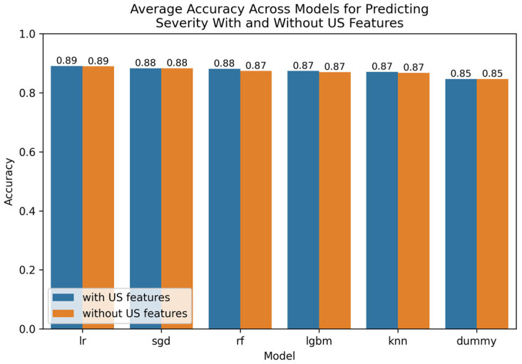
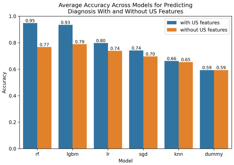
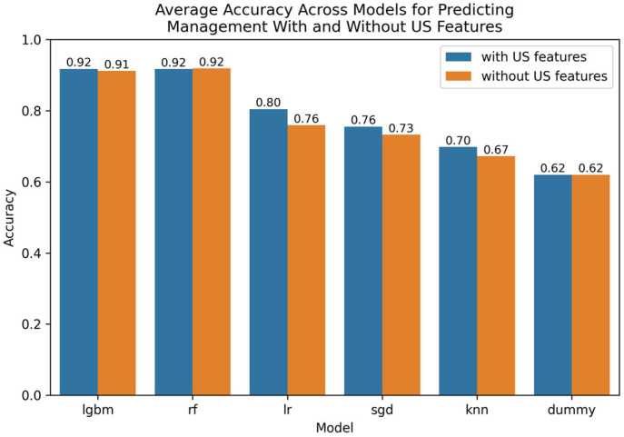
Results: US features significantly improved diagnostic accuracy, supporting their use in reducing model bias. However, they were not essential for maximizing accuracy in predicting management or severity. In summary, our best-performing models were, for diagnosis, the random forest with embedded LGBM feature selection (98.1% accuracy, AUROC: 0.993), for management, the random forest without feature selection (93.9% accuracy, AUROC: 0.980), and for severity, the LGBM with filter-based association feature selection (90.1% accuracy, AUROC: 0.931).
Conclusions: Our results demonstrate that high-performing, interpretable machine learning models can predict key clinical outcomes in pediatric appendicitis. US image features improve diagnostic accuracy but are not critical for predicting management or severity.
TomographyMedicine-Radiology, Nuclear Medicine and Imaging
CiteScore
2.70
自引率
10.50%
发文量
222
期刊介绍:
TomographyTM publishes basic (technical and pre-clinical) and clinical scientific articles which involve the advancement of imaging technologies. Tomography encompasses studies that use single or multiple imaging modalities including for example CT, US, PET, SPECT, MR and hyperpolarization technologies, as well as optical modalities (i.e. bioluminescence, photoacoustic, endomicroscopy, fiber optic imaging and optical computed tomography) in basic sciences, engineering, preclinical and clinical medicine.
Tomography also welcomes studies involving exploration and refinement of contrast mechanisms and image-derived metrics within and across modalities toward the development of novel imaging probes for image-based feedback and intervention. The use of imaging in biology and medicine provides unparalleled opportunities to noninvasively interrogate tissues to obtain real-time dynamic and quantitative information required for diagnosis and response to interventions and to follow evolving pathological conditions. As multi-modal studies and the complexities of imaging technologies themselves are ever increasing to provide advanced information to scientists and clinicians.
Tomography provides a unique publication venue allowing investigators the opportunity to more precisely communicate integrated findings related to the diverse and heterogeneous features associated with underlying anatomical, physiological, functional, metabolic and molecular genetic activities of normal and diseased tissue. Thus Tomography publishes peer-reviewed articles which involve the broad use of imaging of any tissue and disease type including both preclinical and clinical investigations. In addition, hardware/software along with chemical and molecular probe advances are welcome as they are deemed to significantly contribute towards the long-term goal of improving the overall impact of imaging on scientific and clinical discovery.

 求助内容:
求助内容: 应助结果提醒方式:


