Diana M Lindquist, Mary Kate Manhard, Joel Levoy, Jonathan R Dillman
{"title":"钠和酰胺质子转移加权磁共振成像方法在轻度脂肪变性肝病中的可行性。","authors":"Diana M Lindquist, Mary Kate Manhard, Joel Levoy, Jonathan R Dillman","doi":"10.3390/tomography11080089","DOIUrl":null,"url":null,"abstract":"<p><p><b>Background/Objectives</b>: Fat and inflammation confound current magnetic resonance imaging (MRI) methods for assessing fibrosis in liver disease. Sodium or amide proton transfer-weighted MRI methods may be more specific for assessing liver fibrosis. The purpose of this study was to determine the feasibility of sodium and amide proton transfer-weighted MRI in individuals with liver disease and to determine if either method correlated with clinical markers of fibrosis. <b>Methods</b>: T<sub>1</sub> and T<sub>2</sub> relaxation maps, proton density fat fraction maps, liver shear stiffness maps, amide proton transfer-weighted (APTw) images, and sodium images were acquired at 3T. Image data were extracted from regions of interest placed in the liver. ANOVA tests were run with disease status, age, and body mass index as independent factors; significance was set to <i>p</i> < 0.05. Post-hoc t-tests were run when the ANOVA showed significance. <b>Results</b>: A total of 36 participants were enrolled, 34 of whom were included in the final APTw analysis and 24 in the sodium analysis. Estimated liver tissue sodium concentration differentiated participants with liver disease from those without, whereas amide proton transfer-weighted MRI did not. Estimated liver tissue sodium concentration negatively correlated with the Fibrosis-4 score, but amide proton transfer-weighted MRI did not correlate with any clinical marker of disease. <b>Conclusions</b>: Amide proton-weighted imaging was not different between groups. Estimated liver tissue sodium concentrations did differ between groups but did not provide additional information over conventional methods.</p>","PeriodicalId":51330,"journal":{"name":"Tomography","volume":"11 8","pages":""},"PeriodicalIF":2.2000,"publicationDate":"2025-08-06","publicationTypes":"Journal Article","fieldsOfStudy":null,"isOpenAccess":false,"openAccessPdf":"https://www.ncbi.nlm.nih.gov/pmc/articles/PMC12389949/pdf/","citationCount":"0","resultStr":"{\"title\":\"Feasibility of Sodium and Amide Proton Transfer-Weighted Magnetic Resonance Imaging Methods in Mild Steatotic Liver Disease.\",\"authors\":\"Diana M Lindquist, Mary Kate Manhard, Joel Levoy, Jonathan R Dillman\",\"doi\":\"10.3390/tomography11080089\",\"DOIUrl\":null,\"url\":null,\"abstract\":\"<p><p><b>Background/Objectives</b>: Fat and inflammation confound current magnetic resonance imaging (MRI) methods for assessing fibrosis in liver disease. Sodium or amide proton transfer-weighted MRI methods may be more specific for assessing liver fibrosis. The purpose of this study was to determine the feasibility of sodium and amide proton transfer-weighted MRI in individuals with liver disease and to determine if either method correlated with clinical markers of fibrosis. <b>Methods</b>: T<sub>1</sub> and T<sub>2</sub> relaxation maps, proton density fat fraction maps, liver shear stiffness maps, amide proton transfer-weighted (APTw) images, and sodium images were acquired at 3T. Image data were extracted from regions of interest placed in the liver. ANOVA tests were run with disease status, age, and body mass index as independent factors; significance was set to <i>p</i> < 0.05. Post-hoc t-tests were run when the ANOVA showed significance. <b>Results</b>: A total of 36 participants were enrolled, 34 of whom were included in the final APTw analysis and 24 in the sodium analysis. Estimated liver tissue sodium concentration differentiated participants with liver disease from those without, whereas amide proton transfer-weighted MRI did not. Estimated liver tissue sodium concentration negatively correlated with the Fibrosis-4 score, but amide proton transfer-weighted MRI did not correlate with any clinical marker of disease. <b>Conclusions</b>: Amide proton-weighted imaging was not different between groups. Estimated liver tissue sodium concentrations did differ between groups but did not provide additional information over conventional methods.</p>\",\"PeriodicalId\":51330,\"journal\":{\"name\":\"Tomography\",\"volume\":\"11 8\",\"pages\":\"\"},\"PeriodicalIF\":2.2000,\"publicationDate\":\"2025-08-06\",\"publicationTypes\":\"Journal Article\",\"fieldsOfStudy\":null,\"isOpenAccess\":false,\"openAccessPdf\":\"https://www.ncbi.nlm.nih.gov/pmc/articles/PMC12389949/pdf/\",\"citationCount\":\"0\",\"resultStr\":null,\"platform\":\"Semanticscholar\",\"paperid\":null,\"PeriodicalName\":\"Tomography\",\"FirstCategoryId\":\"3\",\"ListUrlMain\":\"https://doi.org/10.3390/tomography11080089\",\"RegionNum\":4,\"RegionCategory\":\"医学\",\"ArticlePicture\":[],\"TitleCN\":null,\"AbstractTextCN\":null,\"PMCID\":null,\"EPubDate\":\"\",\"PubModel\":\"\",\"JCR\":\"Q2\",\"JCRName\":\"RADIOLOGY, NUCLEAR MEDICINE & MEDICAL IMAGING\",\"Score\":null,\"Total\":0}","platform":"Semanticscholar","paperid":null,"PeriodicalName":"Tomography","FirstCategoryId":"3","ListUrlMain":"https://doi.org/10.3390/tomography11080089","RegionNum":4,"RegionCategory":"医学","ArticlePicture":[],"TitleCN":null,"AbstractTextCN":null,"PMCID":null,"EPubDate":"","PubModel":"","JCR":"Q2","JCRName":"RADIOLOGY, NUCLEAR MEDICINE & MEDICAL IMAGING","Score":null,"Total":0}
Feasibility of Sodium and Amide Proton Transfer-Weighted Magnetic Resonance Imaging Methods in Mild Steatotic Liver Disease.
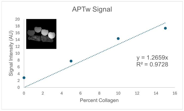
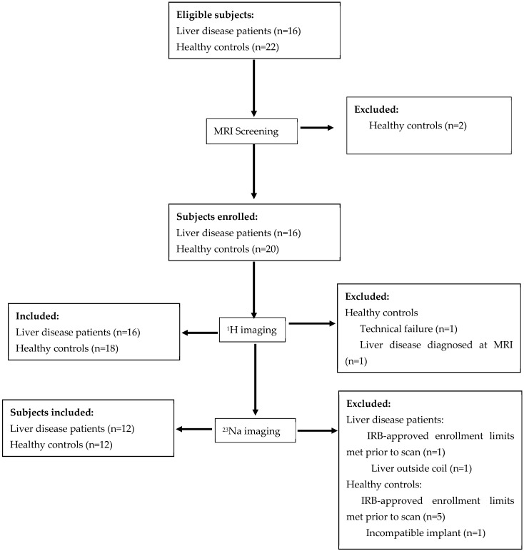
Background/Objectives: Fat and inflammation confound current magnetic resonance imaging (MRI) methods for assessing fibrosis in liver disease. Sodium or amide proton transfer-weighted MRI methods may be more specific for assessing liver fibrosis. The purpose of this study was to determine the feasibility of sodium and amide proton transfer-weighted MRI in individuals with liver disease and to determine if either method correlated with clinical markers of fibrosis. Methods: T1 and T2 relaxation maps, proton density fat fraction maps, liver shear stiffness maps, amide proton transfer-weighted (APTw) images, and sodium images were acquired at 3T. Image data were extracted from regions of interest placed in the liver. ANOVA tests were run with disease status, age, and body mass index as independent factors; significance was set to p < 0.05. Post-hoc t-tests were run when the ANOVA showed significance. Results: A total of 36 participants were enrolled, 34 of whom were included in the final APTw analysis and 24 in the sodium analysis. Estimated liver tissue sodium concentration differentiated participants with liver disease from those without, whereas amide proton transfer-weighted MRI did not. Estimated liver tissue sodium concentration negatively correlated with the Fibrosis-4 score, but amide proton transfer-weighted MRI did not correlate with any clinical marker of disease. Conclusions: Amide proton-weighted imaging was not different between groups. Estimated liver tissue sodium concentrations did differ between groups but did not provide additional information over conventional methods.
TomographyMedicine-Radiology, Nuclear Medicine and Imaging
CiteScore
2.70
自引率
10.50%
发文量
222
期刊介绍:
TomographyTM publishes basic (technical and pre-clinical) and clinical scientific articles which involve the advancement of imaging technologies. Tomography encompasses studies that use single or multiple imaging modalities including for example CT, US, PET, SPECT, MR and hyperpolarization technologies, as well as optical modalities (i.e. bioluminescence, photoacoustic, endomicroscopy, fiber optic imaging and optical computed tomography) in basic sciences, engineering, preclinical and clinical medicine.
Tomography also welcomes studies involving exploration and refinement of contrast mechanisms and image-derived metrics within and across modalities toward the development of novel imaging probes for image-based feedback and intervention. The use of imaging in biology and medicine provides unparalleled opportunities to noninvasively interrogate tissues to obtain real-time dynamic and quantitative information required for diagnosis and response to interventions and to follow evolving pathological conditions. As multi-modal studies and the complexities of imaging technologies themselves are ever increasing to provide advanced information to scientists and clinicians.
Tomography provides a unique publication venue allowing investigators the opportunity to more precisely communicate integrated findings related to the diverse and heterogeneous features associated with underlying anatomical, physiological, functional, metabolic and molecular genetic activities of normal and diseased tissue. Thus Tomography publishes peer-reviewed articles which involve the broad use of imaging of any tissue and disease type including both preclinical and clinical investigations. In addition, hardware/software along with chemical and molecular probe advances are welcome as they are deemed to significantly contribute towards the long-term goal of improving the overall impact of imaging on scientific and clinical discovery.

 求助内容:
求助内容: 应助结果提醒方式:


