Graziella Di Grezia, Sara Mercogliano, Luca Marinelli, Antonio Nazzaro, Alessandro Galiano, Elisa Cisternino, Gianluca Gatta, Vincenzo Cuccurullo, Mariano Scaglione
{"title":"对比增强乳房x光检查在乳腺病变评估中的准确性和手术影响。","authors":"Graziella Di Grezia, Sara Mercogliano, Luca Marinelli, Antonio Nazzaro, Alessandro Galiano, Elisa Cisternino, Gianluca Gatta, Vincenzo Cuccurullo, Mariano Scaglione","doi":"10.3390/tomography11080093","DOIUrl":null,"url":null,"abstract":"<p><strong>Background: </strong>Accurate preoperative tumor sizing is critical for optimal surgical planning in breast cancer. Contrast-enhanced mammography (CEM) has emerged as a promising modality, yet its accuracy relative to conventional imaging and pathology requires further validation.</p><p><strong>Objective: </strong>To prospectively evaluate the dimensional accuracy and reproducibility of CEM compared to mammography and ultrasound, using surgical pathology as the reference standard.</p><p><strong>Methods: </strong>A total of 205 patients with 267 breast lesions underwent preoperative CEM, mammography, and ultrasound. Tumor sizes were measured independently by two radiologists. Accuracy was assessed via mean absolute error (MAE), Pearson and Spearman correlations, and inter-reader agreement evaluated by intraclass correlation coefficient (ICC) and Gwet's AC1. Sensitivity analyses included bootstrap confidence intervals and log-transformed data. The surgical impact of additional lesions detected by CEM was also analyzed.</p><p><strong>Results: </strong>CEM showed superior accuracy with a mean absolute error of 0.46 mm (95% CI: 0.24-0.68) compared to mammography (4.06 mm) and ultrasound (3.52 mm) (<i>p</i> < 0.00001). Pearson's correlation between CEM and pathology was exceptionally high (r = 0.995; 95% CI: 0.994-0.996), with similar robustness after log transformation. Inter-reader agreement for CEM was excellent (ICC 0.93; Gwet's AC1 ~0.96, 95% CI: 0.93-0.98). CEM detected additional lesions in 13.1% of patients, leading to altered surgical management in 6.4%. Background parenchymal enhancement was independently associated with measurement error.</p><p><strong>Conclusions: </strong>CEM provides highly accurate and reproducible tumor size estimation superior to conventional imaging modalities, with potential clinical impact through detection of additional lesions. Its ability to detect additional lesions not seen on mammography or ultrasound has direct implications for surgical decision making, with the potential to reduce reoperations and improve oncologic and cosmetic outcomes. However, high correlation values and selective patient cohorts warrant cautious interpretation. Further multicenter studies are needed to confirm these findings and define CEM's role in clinical practice.</p>","PeriodicalId":51330,"journal":{"name":"Tomography","volume":"11 8","pages":""},"PeriodicalIF":2.2000,"publicationDate":"2025-08-20","publicationTypes":"Journal Article","fieldsOfStudy":null,"isOpenAccess":false,"openAccessPdf":"https://www.ncbi.nlm.nih.gov/pmc/articles/PMC12389778/pdf/","citationCount":"0","resultStr":"{\"title\":\"Contrast-Enhanced Mammography in Breast Lesion Assessment: Accuracy and Surgical Impact.\",\"authors\":\"Graziella Di Grezia, Sara Mercogliano, Luca Marinelli, Antonio Nazzaro, Alessandro Galiano, Elisa Cisternino, Gianluca Gatta, Vincenzo Cuccurullo, Mariano Scaglione\",\"doi\":\"10.3390/tomography11080093\",\"DOIUrl\":null,\"url\":null,\"abstract\":\"<p><strong>Background: </strong>Accurate preoperative tumor sizing is critical for optimal surgical planning in breast cancer. Contrast-enhanced mammography (CEM) has emerged as a promising modality, yet its accuracy relative to conventional imaging and pathology requires further validation.</p><p><strong>Objective: </strong>To prospectively evaluate the dimensional accuracy and reproducibility of CEM compared to mammography and ultrasound, using surgical pathology as the reference standard.</p><p><strong>Methods: </strong>A total of 205 patients with 267 breast lesions underwent preoperative CEM, mammography, and ultrasound. Tumor sizes were measured independently by two radiologists. Accuracy was assessed via mean absolute error (MAE), Pearson and Spearman correlations, and inter-reader agreement evaluated by intraclass correlation coefficient (ICC) and Gwet's AC1. Sensitivity analyses included bootstrap confidence intervals and log-transformed data. The surgical impact of additional lesions detected by CEM was also analyzed.</p><p><strong>Results: </strong>CEM showed superior accuracy with a mean absolute error of 0.46 mm (95% CI: 0.24-0.68) compared to mammography (4.06 mm) and ultrasound (3.52 mm) (<i>p</i> < 0.00001). Pearson's correlation between CEM and pathology was exceptionally high (r = 0.995; 95% CI: 0.994-0.996), with similar robustness after log transformation. Inter-reader agreement for CEM was excellent (ICC 0.93; Gwet's AC1 ~0.96, 95% CI: 0.93-0.98). CEM detected additional lesions in 13.1% of patients, leading to altered surgical management in 6.4%. Background parenchymal enhancement was independently associated with measurement error.</p><p><strong>Conclusions: </strong>CEM provides highly accurate and reproducible tumor size estimation superior to conventional imaging modalities, with potential clinical impact through detection of additional lesions. Its ability to detect additional lesions not seen on mammography or ultrasound has direct implications for surgical decision making, with the potential to reduce reoperations and improve oncologic and cosmetic outcomes. However, high correlation values and selective patient cohorts warrant cautious interpretation. Further multicenter studies are needed to confirm these findings and define CEM's role in clinical practice.</p>\",\"PeriodicalId\":51330,\"journal\":{\"name\":\"Tomography\",\"volume\":\"11 8\",\"pages\":\"\"},\"PeriodicalIF\":2.2000,\"publicationDate\":\"2025-08-20\",\"publicationTypes\":\"Journal Article\",\"fieldsOfStudy\":null,\"isOpenAccess\":false,\"openAccessPdf\":\"https://www.ncbi.nlm.nih.gov/pmc/articles/PMC12389778/pdf/\",\"citationCount\":\"0\",\"resultStr\":null,\"platform\":\"Semanticscholar\",\"paperid\":null,\"PeriodicalName\":\"Tomography\",\"FirstCategoryId\":\"3\",\"ListUrlMain\":\"https://doi.org/10.3390/tomography11080093\",\"RegionNum\":4,\"RegionCategory\":\"医学\",\"ArticlePicture\":[],\"TitleCN\":null,\"AbstractTextCN\":null,\"PMCID\":null,\"EPubDate\":\"\",\"PubModel\":\"\",\"JCR\":\"Q2\",\"JCRName\":\"RADIOLOGY, NUCLEAR MEDICINE & MEDICAL IMAGING\",\"Score\":null,\"Total\":0}","platform":"Semanticscholar","paperid":null,"PeriodicalName":"Tomography","FirstCategoryId":"3","ListUrlMain":"https://doi.org/10.3390/tomography11080093","RegionNum":4,"RegionCategory":"医学","ArticlePicture":[],"TitleCN":null,"AbstractTextCN":null,"PMCID":null,"EPubDate":"","PubModel":"","JCR":"Q2","JCRName":"RADIOLOGY, NUCLEAR MEDICINE & MEDICAL IMAGING","Score":null,"Total":0}
Contrast-Enhanced Mammography in Breast Lesion Assessment: Accuracy and Surgical Impact.
Background: Accurate preoperative tumor sizing is critical for optimal surgical planning in breast cancer. Contrast-enhanced mammography (CEM) has emerged as a promising modality, yet its accuracy relative to conventional imaging and pathology requires further validation.
Objective: To prospectively evaluate the dimensional accuracy and reproducibility of CEM compared to mammography and ultrasound, using surgical pathology as the reference standard.
Methods: A total of 205 patients with 267 breast lesions underwent preoperative CEM, mammography, and ultrasound. Tumor sizes were measured independently by two radiologists. Accuracy was assessed via mean absolute error (MAE), Pearson and Spearman correlations, and inter-reader agreement evaluated by intraclass correlation coefficient (ICC) and Gwet's AC1. Sensitivity analyses included bootstrap confidence intervals and log-transformed data. The surgical impact of additional lesions detected by CEM was also analyzed.
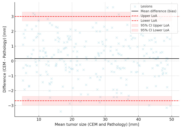
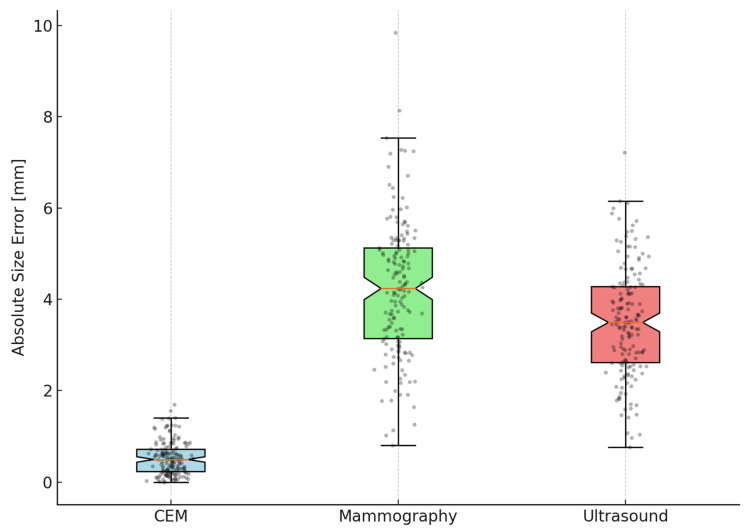
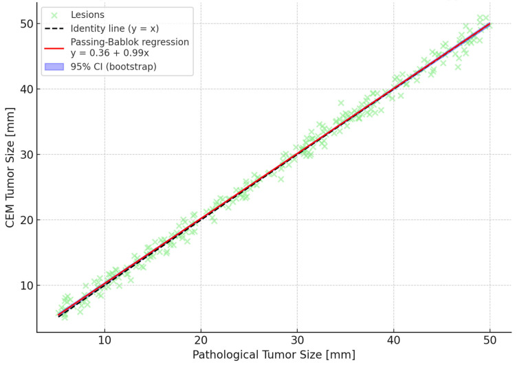
Results: CEM showed superior accuracy with a mean absolute error of 0.46 mm (95% CI: 0.24-0.68) compared to mammography (4.06 mm) and ultrasound (3.52 mm) (p < 0.00001). Pearson's correlation between CEM and pathology was exceptionally high (r = 0.995; 95% CI: 0.994-0.996), with similar robustness after log transformation. Inter-reader agreement for CEM was excellent (ICC 0.93; Gwet's AC1 ~0.96, 95% CI: 0.93-0.98). CEM detected additional lesions in 13.1% of patients, leading to altered surgical management in 6.4%. Background parenchymal enhancement was independently associated with measurement error.
Conclusions: CEM provides highly accurate and reproducible tumor size estimation superior to conventional imaging modalities, with potential clinical impact through detection of additional lesions. Its ability to detect additional lesions not seen on mammography or ultrasound has direct implications for surgical decision making, with the potential to reduce reoperations and improve oncologic and cosmetic outcomes. However, high correlation values and selective patient cohorts warrant cautious interpretation. Further multicenter studies are needed to confirm these findings and define CEM's role in clinical practice.
TomographyMedicine-Radiology, Nuclear Medicine and Imaging
CiteScore
2.70
自引率
10.50%
发文量
222
期刊介绍:
TomographyTM publishes basic (technical and pre-clinical) and clinical scientific articles which involve the advancement of imaging technologies. Tomography encompasses studies that use single or multiple imaging modalities including for example CT, US, PET, SPECT, MR and hyperpolarization technologies, as well as optical modalities (i.e. bioluminescence, photoacoustic, endomicroscopy, fiber optic imaging and optical computed tomography) in basic sciences, engineering, preclinical and clinical medicine.
Tomography also welcomes studies involving exploration and refinement of contrast mechanisms and image-derived metrics within and across modalities toward the development of novel imaging probes for image-based feedback and intervention. The use of imaging in biology and medicine provides unparalleled opportunities to noninvasively interrogate tissues to obtain real-time dynamic and quantitative information required for diagnosis and response to interventions and to follow evolving pathological conditions. As multi-modal studies and the complexities of imaging technologies themselves are ever increasing to provide advanced information to scientists and clinicians.
Tomography provides a unique publication venue allowing investigators the opportunity to more precisely communicate integrated findings related to the diverse and heterogeneous features associated with underlying anatomical, physiological, functional, metabolic and molecular genetic activities of normal and diseased tissue. Thus Tomography publishes peer-reviewed articles which involve the broad use of imaging of any tissue and disease type including both preclinical and clinical investigations. In addition, hardware/software along with chemical and molecular probe advances are welcome as they are deemed to significantly contribute towards the long-term goal of improving the overall impact of imaging on scientific and clinical discovery.

 求助内容:
求助内容: 应助结果提醒方式:


