Justine Schoch, Viola Düring, Michael Wiedmann, Daniel Overhoff, Daniel Dillinger, Stephan Waldeck, Hans-Ulrich Schmelz, Tim Nestler
{"title":"间隔6周mpMRI扫描在前列腺癌PI-RADS分类中的差异","authors":"Justine Schoch, Viola Düring, Michael Wiedmann, Daniel Overhoff, Daniel Dillinger, Stephan Waldeck, Hans-Ulrich Schmelz, Tim Nestler","doi":"10.3390/tomography11080092","DOIUrl":null,"url":null,"abstract":"<p><strong>Objectives: </strong>This study aimed to investigate the consistency of lesion identification by Prostate Imaging Reporting and Data System (PI-RADS) and the related clinical and histological characteristics in a high-volume tertiary care center.</p><p><strong>Materials and methods: </strong>The analysis used real-world data from 111 patients between 2018 and 2022. Each patient underwent two multiparametric magnetic resonance imaging (MRI) scans of the prostate at different institutions with a median interval of 42 days between the scans, followed by an MRI-fused biopsy conducted 7 days after the second MRI.</p><p><strong>Results: </strong>The PI-RADS classifications assigned to the index lesions in the in-house prostate MRI were as follows: PI-RADS V, 33.3% (n = 37); PI-RADS IV, 49.5% (n = 55); PI-RADS III, 12.6% (n = 14); and PI-RADS II, 4.5% (n = 5). Cancer detection rates for randomized and/or targeted biopsies were 91.9% (n = 34) for PI-RADS V, 65.5% (n = 36) for PI-RADS IV, 21.4% (n = 3) for PI-RADS III, and 20% (n = 1) for PI-RADS II. Overall, malignant histology was observed in 64.9% (n = 72) of the targeted lesions and 57.7% (n = 64) of the randomized biopsies. In the first performed, external MRI, 18% (n = 20) and 10.8% (n = 12) of the patients were classified in the higher and lower PI-RADS categories, respectively. The biopsy plan was adjusted for 57 patients (51.4%); nevertheless, any cancer could have possibly been identified regardless of the adjustments.</p><p><strong>Conclusion: </strong>The 6-week interval between the MRI scans did not affect the quality of the biopsy results significantly.</p>","PeriodicalId":51330,"journal":{"name":"Tomography","volume":"11 8","pages":""},"PeriodicalIF":2.2000,"publicationDate":"2025-08-18","publicationTypes":"Journal Article","fieldsOfStudy":null,"isOpenAccess":false,"openAccessPdf":"https://www.ncbi.nlm.nih.gov/pmc/articles/PMC12390117/pdf/","citationCount":"0","resultStr":"{\"title\":\"Differences in PI-RADS Classification of Prostate Cancer Based on mpMRI Scans Taken 6 Weeks Apart.\",\"authors\":\"Justine Schoch, Viola Düring, Michael Wiedmann, Daniel Overhoff, Daniel Dillinger, Stephan Waldeck, Hans-Ulrich Schmelz, Tim Nestler\",\"doi\":\"10.3390/tomography11080092\",\"DOIUrl\":null,\"url\":null,\"abstract\":\"<p><strong>Objectives: </strong>This study aimed to investigate the consistency of lesion identification by Prostate Imaging Reporting and Data System (PI-RADS) and the related clinical and histological characteristics in a high-volume tertiary care center.</p><p><strong>Materials and methods: </strong>The analysis used real-world data from 111 patients between 2018 and 2022. Each patient underwent two multiparametric magnetic resonance imaging (MRI) scans of the prostate at different institutions with a median interval of 42 days between the scans, followed by an MRI-fused biopsy conducted 7 days after the second MRI.</p><p><strong>Results: </strong>The PI-RADS classifications assigned to the index lesions in the in-house prostate MRI were as follows: PI-RADS V, 33.3% (n = 37); PI-RADS IV, 49.5% (n = 55); PI-RADS III, 12.6% (n = 14); and PI-RADS II, 4.5% (n = 5). Cancer detection rates for randomized and/or targeted biopsies were 91.9% (n = 34) for PI-RADS V, 65.5% (n = 36) for PI-RADS IV, 21.4% (n = 3) for PI-RADS III, and 20% (n = 1) for PI-RADS II. Overall, malignant histology was observed in 64.9% (n = 72) of the targeted lesions and 57.7% (n = 64) of the randomized biopsies. In the first performed, external MRI, 18% (n = 20) and 10.8% (n = 12) of the patients were classified in the higher and lower PI-RADS categories, respectively. The biopsy plan was adjusted for 57 patients (51.4%); nevertheless, any cancer could have possibly been identified regardless of the adjustments.</p><p><strong>Conclusion: </strong>The 6-week interval between the MRI scans did not affect the quality of the biopsy results significantly.</p>\",\"PeriodicalId\":51330,\"journal\":{\"name\":\"Tomography\",\"volume\":\"11 8\",\"pages\":\"\"},\"PeriodicalIF\":2.2000,\"publicationDate\":\"2025-08-18\",\"publicationTypes\":\"Journal Article\",\"fieldsOfStudy\":null,\"isOpenAccess\":false,\"openAccessPdf\":\"https://www.ncbi.nlm.nih.gov/pmc/articles/PMC12390117/pdf/\",\"citationCount\":\"0\",\"resultStr\":null,\"platform\":\"Semanticscholar\",\"paperid\":null,\"PeriodicalName\":\"Tomography\",\"FirstCategoryId\":\"3\",\"ListUrlMain\":\"https://doi.org/10.3390/tomography11080092\",\"RegionNum\":4,\"RegionCategory\":\"医学\",\"ArticlePicture\":[],\"TitleCN\":null,\"AbstractTextCN\":null,\"PMCID\":null,\"EPubDate\":\"\",\"PubModel\":\"\",\"JCR\":\"Q2\",\"JCRName\":\"RADIOLOGY, NUCLEAR MEDICINE & MEDICAL IMAGING\",\"Score\":null,\"Total\":0}","platform":"Semanticscholar","paperid":null,"PeriodicalName":"Tomography","FirstCategoryId":"3","ListUrlMain":"https://doi.org/10.3390/tomography11080092","RegionNum":4,"RegionCategory":"医学","ArticlePicture":[],"TitleCN":null,"AbstractTextCN":null,"PMCID":null,"EPubDate":"","PubModel":"","JCR":"Q2","JCRName":"RADIOLOGY, NUCLEAR MEDICINE & MEDICAL IMAGING","Score":null,"Total":0}
Differences in PI-RADS Classification of Prostate Cancer Based on mpMRI Scans Taken 6 Weeks Apart.
Objectives: This study aimed to investigate the consistency of lesion identification by Prostate Imaging Reporting and Data System (PI-RADS) and the related clinical and histological characteristics in a high-volume tertiary care center.
Materials and methods: The analysis used real-world data from 111 patients between 2018 and 2022. Each patient underwent two multiparametric magnetic resonance imaging (MRI) scans of the prostate at different institutions with a median interval of 42 days between the scans, followed by an MRI-fused biopsy conducted 7 days after the second MRI.
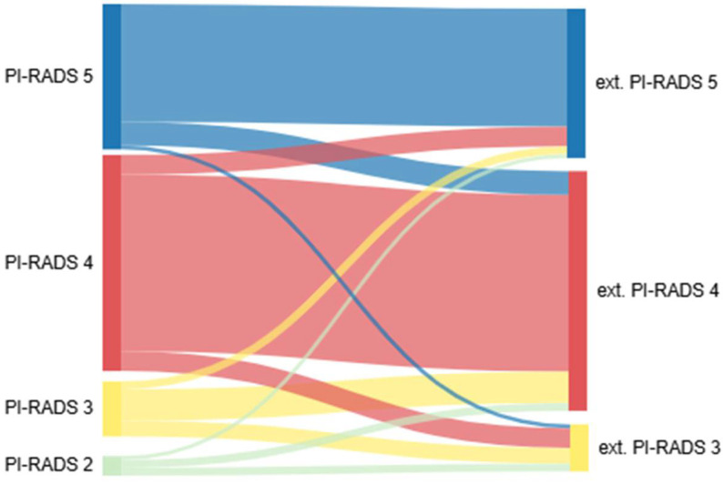
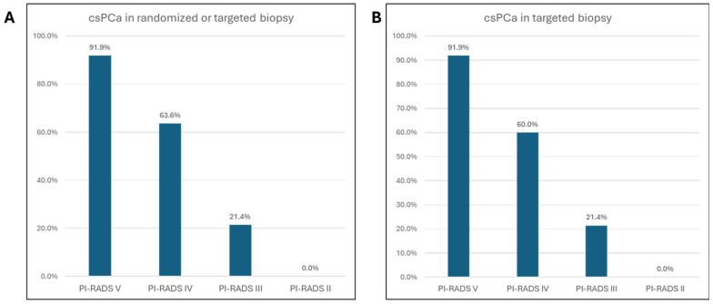
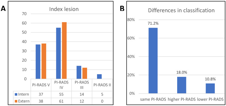
Results: The PI-RADS classifications assigned to the index lesions in the in-house prostate MRI were as follows: PI-RADS V, 33.3% (n = 37); PI-RADS IV, 49.5% (n = 55); PI-RADS III, 12.6% (n = 14); and PI-RADS II, 4.5% (n = 5). Cancer detection rates for randomized and/or targeted biopsies were 91.9% (n = 34) for PI-RADS V, 65.5% (n = 36) for PI-RADS IV, 21.4% (n = 3) for PI-RADS III, and 20% (n = 1) for PI-RADS II. Overall, malignant histology was observed in 64.9% (n = 72) of the targeted lesions and 57.7% (n = 64) of the randomized biopsies. In the first performed, external MRI, 18% (n = 20) and 10.8% (n = 12) of the patients were classified in the higher and lower PI-RADS categories, respectively. The biopsy plan was adjusted for 57 patients (51.4%); nevertheless, any cancer could have possibly been identified regardless of the adjustments.
Conclusion: The 6-week interval between the MRI scans did not affect the quality of the biopsy results significantly.
TomographyMedicine-Radiology, Nuclear Medicine and Imaging
CiteScore
2.70
自引率
10.50%
发文量
222
期刊介绍:
TomographyTM publishes basic (technical and pre-clinical) and clinical scientific articles which involve the advancement of imaging technologies. Tomography encompasses studies that use single or multiple imaging modalities including for example CT, US, PET, SPECT, MR and hyperpolarization technologies, as well as optical modalities (i.e. bioluminescence, photoacoustic, endomicroscopy, fiber optic imaging and optical computed tomography) in basic sciences, engineering, preclinical and clinical medicine.
Tomography also welcomes studies involving exploration and refinement of contrast mechanisms and image-derived metrics within and across modalities toward the development of novel imaging probes for image-based feedback and intervention. The use of imaging in biology and medicine provides unparalleled opportunities to noninvasively interrogate tissues to obtain real-time dynamic and quantitative information required for diagnosis and response to interventions and to follow evolving pathological conditions. As multi-modal studies and the complexities of imaging technologies themselves are ever increasing to provide advanced information to scientists and clinicians.
Tomography provides a unique publication venue allowing investigators the opportunity to more precisely communicate integrated findings related to the diverse and heterogeneous features associated with underlying anatomical, physiological, functional, metabolic and molecular genetic activities of normal and diseased tissue. Thus Tomography publishes peer-reviewed articles which involve the broad use of imaging of any tissue and disease type including both preclinical and clinical investigations. In addition, hardware/software along with chemical and molecular probe advances are welcome as they are deemed to significantly contribute towards the long-term goal of improving the overall impact of imaging on scientific and clinical discovery.

 求助内容:
求助内容: 应助结果提醒方式:


