Hashim Alhammouri, Ramzi Ibrahim, Rahmeh Alasmar, Mahmoud Abdelnabi, Eiad Habib, Mohamed Allam, Hoang Nhat Pham, Hossam Elbenawi, Juan Farina, Balaji Tamarappoo, Clinton Jokerst, Kwan Lee, Chadi Ayoub, Reza Arsanjani
{"title":"计算机断层扫描和冠状动脉斑块分析。","authors":"Hashim Alhammouri, Ramzi Ibrahim, Rahmeh Alasmar, Mahmoud Abdelnabi, Eiad Habib, Mohamed Allam, Hoang Nhat Pham, Hossam Elbenawi, Juan Farina, Balaji Tamarappoo, Clinton Jokerst, Kwan Lee, Chadi Ayoub, Reza Arsanjani","doi":"10.3390/tomography11080085","DOIUrl":null,"url":null,"abstract":"<p><p>Advances in plaque imaging have transformed cardiovascular diagnostics through detailed characterization of atherosclerotic plaques beyond traditional stenosis assessment. This review outlines the clinical applications of varying modalities, including dual-layer spectral CT, photon-counting CT, dual-energy CT, and CT-derived fractional flow reserve (CT-FFR). These technologies offer improved spatial resolution, tissue differentiation, and functional assessment of coronary lesions. Additionally, artificial intelligence has emerged as a powerful tool to automate plaque detection, quantify burden, and refine risk prediction. Collectively, these innovations provide a more comprehensive approach to coronary artery disease evaluation and support personalized management strategies.</p>","PeriodicalId":51330,"journal":{"name":"Tomography","volume":"11 8","pages":""},"PeriodicalIF":2.2000,"publicationDate":"2025-07-30","publicationTypes":"Journal Article","fieldsOfStudy":null,"isOpenAccess":false,"openAccessPdf":"https://www.ncbi.nlm.nih.gov/pmc/articles/PMC12389945/pdf/","citationCount":"0","resultStr":"{\"title\":\"Computed Tomography and Coronary Plaque Analysis.\",\"authors\":\"Hashim Alhammouri, Ramzi Ibrahim, Rahmeh Alasmar, Mahmoud Abdelnabi, Eiad Habib, Mohamed Allam, Hoang Nhat Pham, Hossam Elbenawi, Juan Farina, Balaji Tamarappoo, Clinton Jokerst, Kwan Lee, Chadi Ayoub, Reza Arsanjani\",\"doi\":\"10.3390/tomography11080085\",\"DOIUrl\":null,\"url\":null,\"abstract\":\"<p><p>Advances in plaque imaging have transformed cardiovascular diagnostics through detailed characterization of atherosclerotic plaques beyond traditional stenosis assessment. This review outlines the clinical applications of varying modalities, including dual-layer spectral CT, photon-counting CT, dual-energy CT, and CT-derived fractional flow reserve (CT-FFR). These technologies offer improved spatial resolution, tissue differentiation, and functional assessment of coronary lesions. Additionally, artificial intelligence has emerged as a powerful tool to automate plaque detection, quantify burden, and refine risk prediction. Collectively, these innovations provide a more comprehensive approach to coronary artery disease evaluation and support personalized management strategies.</p>\",\"PeriodicalId\":51330,\"journal\":{\"name\":\"Tomography\",\"volume\":\"11 8\",\"pages\":\"\"},\"PeriodicalIF\":2.2000,\"publicationDate\":\"2025-07-30\",\"publicationTypes\":\"Journal Article\",\"fieldsOfStudy\":null,\"isOpenAccess\":false,\"openAccessPdf\":\"https://www.ncbi.nlm.nih.gov/pmc/articles/PMC12389945/pdf/\",\"citationCount\":\"0\",\"resultStr\":null,\"platform\":\"Semanticscholar\",\"paperid\":null,\"PeriodicalName\":\"Tomography\",\"FirstCategoryId\":\"3\",\"ListUrlMain\":\"https://doi.org/10.3390/tomography11080085\",\"RegionNum\":4,\"RegionCategory\":\"医学\",\"ArticlePicture\":[],\"TitleCN\":null,\"AbstractTextCN\":null,\"PMCID\":null,\"EPubDate\":\"\",\"PubModel\":\"\",\"JCR\":\"Q2\",\"JCRName\":\"RADIOLOGY, NUCLEAR MEDICINE & MEDICAL IMAGING\",\"Score\":null,\"Total\":0}","platform":"Semanticscholar","paperid":null,"PeriodicalName":"Tomography","FirstCategoryId":"3","ListUrlMain":"https://doi.org/10.3390/tomography11080085","RegionNum":4,"RegionCategory":"医学","ArticlePicture":[],"TitleCN":null,"AbstractTextCN":null,"PMCID":null,"EPubDate":"","PubModel":"","JCR":"Q2","JCRName":"RADIOLOGY, NUCLEAR MEDICINE & MEDICAL IMAGING","Score":null,"Total":0}
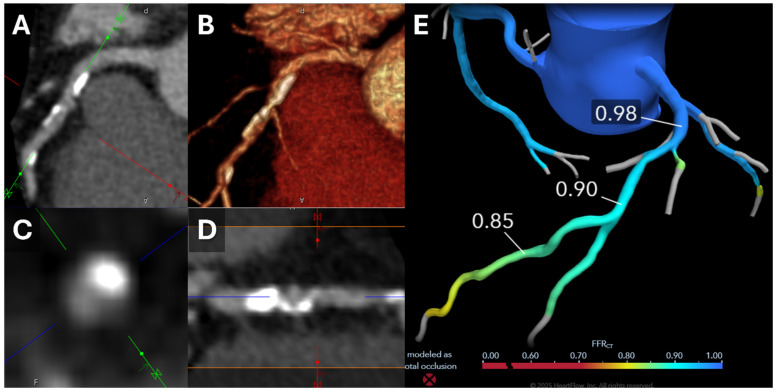
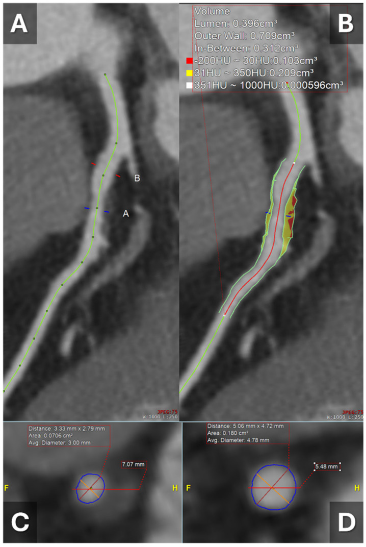
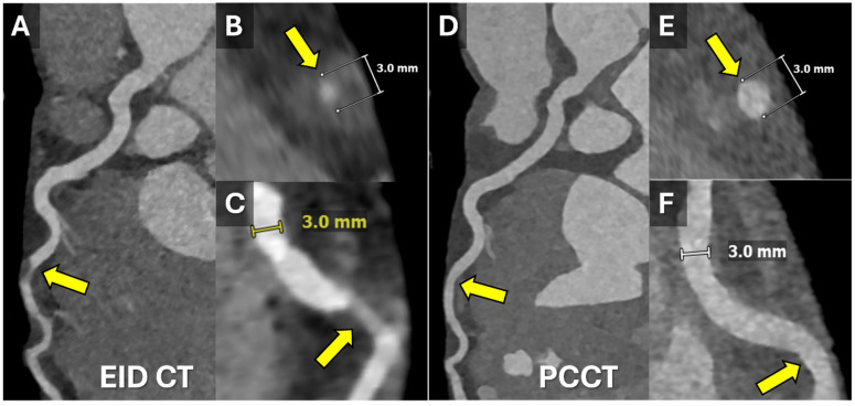
Advances in plaque imaging have transformed cardiovascular diagnostics through detailed characterization of atherosclerotic plaques beyond traditional stenosis assessment. This review outlines the clinical applications of varying modalities, including dual-layer spectral CT, photon-counting CT, dual-energy CT, and CT-derived fractional flow reserve (CT-FFR). These technologies offer improved spatial resolution, tissue differentiation, and functional assessment of coronary lesions. Additionally, artificial intelligence has emerged as a powerful tool to automate plaque detection, quantify burden, and refine risk prediction. Collectively, these innovations provide a more comprehensive approach to coronary artery disease evaluation and support personalized management strategies.
TomographyMedicine-Radiology, Nuclear Medicine and Imaging
CiteScore
2.70
自引率
10.50%
发文量
222
期刊介绍:
TomographyTM publishes basic (technical and pre-clinical) and clinical scientific articles which involve the advancement of imaging technologies. Tomography encompasses studies that use single or multiple imaging modalities including for example CT, US, PET, SPECT, MR and hyperpolarization technologies, as well as optical modalities (i.e. bioluminescence, photoacoustic, endomicroscopy, fiber optic imaging and optical computed tomography) in basic sciences, engineering, preclinical and clinical medicine.
Tomography also welcomes studies involving exploration and refinement of contrast mechanisms and image-derived metrics within and across modalities toward the development of novel imaging probes for image-based feedback and intervention. The use of imaging in biology and medicine provides unparalleled opportunities to noninvasively interrogate tissues to obtain real-time dynamic and quantitative information required for diagnosis and response to interventions and to follow evolving pathological conditions. As multi-modal studies and the complexities of imaging technologies themselves are ever increasing to provide advanced information to scientists and clinicians.
Tomography provides a unique publication venue allowing investigators the opportunity to more precisely communicate integrated findings related to the diverse and heterogeneous features associated with underlying anatomical, physiological, functional, metabolic and molecular genetic activities of normal and diseased tissue. Thus Tomography publishes peer-reviewed articles which involve the broad use of imaging of any tissue and disease type including both preclinical and clinical investigations. In addition, hardware/software along with chemical and molecular probe advances are welcome as they are deemed to significantly contribute towards the long-term goal of improving the overall impact of imaging on scientific and clinical discovery.

 求助内容:
求助内容: 应助结果提醒方式:


