Bano Alsaleh, Ahmed Alanzi, Mohamed Alsaleh, Ahmed Alsaleh, Fouad Aladel
{"title":"外阴皮肤纤维肉瘤隆突,一个不寻常的解剖位置。","authors":"Bano Alsaleh, Ahmed Alanzi, Mohamed Alsaleh, Ahmed Alsaleh, Fouad Aladel","doi":"10.1093/bjrcr/uaaf030","DOIUrl":null,"url":null,"abstract":"<p><p>Vulvar dermatofibrosarcoma protuberans (DFSP) is a rare pathology. So far, only limited number of cases have been reported in literature. In the present case, we discuss a 38-year-old female presented with a painful left vulvar mass. She had a prior history of a left vulvar mass excision which was histopathologically confirmed as benign spindle cell epithelioma. The current mass, extending from the left labia majora to the left gluteal fold, was assessed via contrast-enhanced magnetic resonance imaging (MRI), revealing a well-defined, lobulated lesion with proximity to the distal urethra and clitoris without definite invasion. The patient underwent a wide local excision, radical vulvectomy, and left inguinofemoral lymphadenectomy. Postoperatively, she experienced fever, vulvar swelling, and dysuria. Follow-up MRI demonstrated total resolution of the vulvar mass and collection with no recurrence. Histopathology identified the mass as DFSP, with all surgical margins negative.</p>","PeriodicalId":45216,"journal":{"name":"BJR Case Reports","volume":"11 4","pages":"uaaf030"},"PeriodicalIF":0.5000,"publicationDate":"2025-05-29","publicationTypes":"Journal Article","fieldsOfStudy":null,"isOpenAccess":false,"openAccessPdf":"https://www.ncbi.nlm.nih.gov/pmc/articles/PMC12375401/pdf/","citationCount":"0","resultStr":"{\"title\":\"Vulvar dermatofibrosarcoma protuberans, an unusual anatomical location.\",\"authors\":\"Bano Alsaleh, Ahmed Alanzi, Mohamed Alsaleh, Ahmed Alsaleh, Fouad Aladel\",\"doi\":\"10.1093/bjrcr/uaaf030\",\"DOIUrl\":null,\"url\":null,\"abstract\":\"<p><p>Vulvar dermatofibrosarcoma protuberans (DFSP) is a rare pathology. So far, only limited number of cases have been reported in literature. In the present case, we discuss a 38-year-old female presented with a painful left vulvar mass. She had a prior history of a left vulvar mass excision which was histopathologically confirmed as benign spindle cell epithelioma. The current mass, extending from the left labia majora to the left gluteal fold, was assessed via contrast-enhanced magnetic resonance imaging (MRI), revealing a well-defined, lobulated lesion with proximity to the distal urethra and clitoris without definite invasion. The patient underwent a wide local excision, radical vulvectomy, and left inguinofemoral lymphadenectomy. Postoperatively, she experienced fever, vulvar swelling, and dysuria. Follow-up MRI demonstrated total resolution of the vulvar mass and collection with no recurrence. Histopathology identified the mass as DFSP, with all surgical margins negative.</p>\",\"PeriodicalId\":45216,\"journal\":{\"name\":\"BJR Case Reports\",\"volume\":\"11 4\",\"pages\":\"uaaf030\"},\"PeriodicalIF\":0.5000,\"publicationDate\":\"2025-05-29\",\"publicationTypes\":\"Journal Article\",\"fieldsOfStudy\":null,\"isOpenAccess\":false,\"openAccessPdf\":\"https://www.ncbi.nlm.nih.gov/pmc/articles/PMC12375401/pdf/\",\"citationCount\":\"0\",\"resultStr\":null,\"platform\":\"Semanticscholar\",\"paperid\":null,\"PeriodicalName\":\"BJR Case Reports\",\"FirstCategoryId\":\"1085\",\"ListUrlMain\":\"https://doi.org/10.1093/bjrcr/uaaf030\",\"RegionNum\":0,\"RegionCategory\":null,\"ArticlePicture\":[],\"TitleCN\":null,\"AbstractTextCN\":null,\"PMCID\":null,\"EPubDate\":\"2025/7/1 0:00:00\",\"PubModel\":\"eCollection\",\"JCR\":\"Q4\",\"JCRName\":\"RADIOLOGY, NUCLEAR MEDICINE & MEDICAL IMAGING\",\"Score\":null,\"Total\":0}","platform":"Semanticscholar","paperid":null,"PeriodicalName":"BJR Case Reports","FirstCategoryId":"1085","ListUrlMain":"https://doi.org/10.1093/bjrcr/uaaf030","RegionNum":0,"RegionCategory":null,"ArticlePicture":[],"TitleCN":null,"AbstractTextCN":null,"PMCID":null,"EPubDate":"2025/7/1 0:00:00","PubModel":"eCollection","JCR":"Q4","JCRName":"RADIOLOGY, NUCLEAR MEDICINE & MEDICAL IMAGING","Score":null,"Total":0}
Vulvar dermatofibrosarcoma protuberans, an unusual anatomical location.
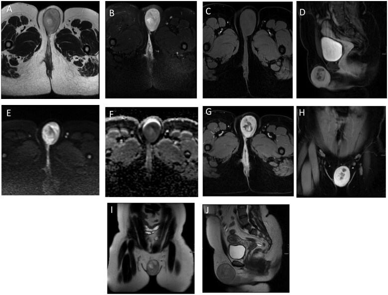
Vulvar dermatofibrosarcoma protuberans (DFSP) is a rare pathology. So far, only limited number of cases have been reported in literature. In the present case, we discuss a 38-year-old female presented with a painful left vulvar mass. She had a prior history of a left vulvar mass excision which was histopathologically confirmed as benign spindle cell epithelioma. The current mass, extending from the left labia majora to the left gluteal fold, was assessed via contrast-enhanced magnetic resonance imaging (MRI), revealing a well-defined, lobulated lesion with proximity to the distal urethra and clitoris without definite invasion. The patient underwent a wide local excision, radical vulvectomy, and left inguinofemoral lymphadenectomy. Postoperatively, she experienced fever, vulvar swelling, and dysuria. Follow-up MRI demonstrated total resolution of the vulvar mass and collection with no recurrence. Histopathology identified the mass as DFSP, with all surgical margins negative.

 求助内容:
求助内容: 应助结果提醒方式:


