A A Musa, R Shamsuddin-Perisamy, C L Low, A H Zulkifly, R Y Kow
{"title":"膝骨关节炎患者仰卧位与站立位膝平片的差异。","authors":"A A Musa, R Shamsuddin-Perisamy, C L Low, A H Zulkifly, R Y Kow","doi":"10.5704/MOJ.2507.007","DOIUrl":null,"url":null,"abstract":"<p><strong>Introduction: </strong>Knee osteoarthritis is a degenerative joint disease attributed to failure in joint repair process. Key aspect of the diagnosis relies on thorough history, along with physical examination and radiology findings. The conventional weight-bearing plain radiograph remains the key modality to determine the severity of the condition and helps to plan the surgery. Nevertheless, not all patients can undergo weight-bearing plain radiographs, especially those who are wheelchair-bound or have severe deformities. The purpose of this study is to investigate whether a weight-bearing plain radiograph of the knee is essential in all patients with knee osteoarthritis.</p><p><strong>Materials and methods: </strong>A prospective cohort study on patients with knee osteoarthritis receiving treatment in a single tertiary hospital was conducted. All patients consented to participate in this study. Patients were assessed functionally with the Western Ontario and McMaster Universities Osteoarthritis Index (WOMAC) and radiologically with plain radiographs. Patients were subjected to undergo both supine and standing plain radiographs of the knee in the same setting for comparison purposes where measurement is done following patient functional outcome and radiological measurement for the patient.</p><p><strong>Results: </strong>Our study shows that reduction in joint space is more obvious in weight-bearing radiographs, however in severe or higher-grade osteoarthritis, a supine radiograph is adequate to diagnose knee osteoarthritis.</p><p><strong>Conclusion: </strong>Standing radiograph of the knee is preferred to a supine knee radiograph wherever possible due to the additional value it brings, however, in certain patient conditions, a supine radiograph is still acceptable.</p>","PeriodicalId":45241,"journal":{"name":"Malaysian Orthopaedic Journal","volume":"19 2","pages":"50-56"},"PeriodicalIF":0.6000,"publicationDate":"2025-07-01","publicationTypes":"Journal Article","fieldsOfStudy":null,"isOpenAccess":false,"openAccessPdf":"https://www.ncbi.nlm.nih.gov/pmc/articles/PMC12368450/pdf/","citationCount":"0","resultStr":"{\"title\":\"The Difference in Supine versus Standing Plain Radiograph of the Knee in Patients with Knee Osteoarthritis.\",\"authors\":\"A A Musa, R Shamsuddin-Perisamy, C L Low, A H Zulkifly, R Y Kow\",\"doi\":\"10.5704/MOJ.2507.007\",\"DOIUrl\":null,\"url\":null,\"abstract\":\"<p><strong>Introduction: </strong>Knee osteoarthritis is a degenerative joint disease attributed to failure in joint repair process. Key aspect of the diagnosis relies on thorough history, along with physical examination and radiology findings. The conventional weight-bearing plain radiograph remains the key modality to determine the severity of the condition and helps to plan the surgery. Nevertheless, not all patients can undergo weight-bearing plain radiographs, especially those who are wheelchair-bound or have severe deformities. The purpose of this study is to investigate whether a weight-bearing plain radiograph of the knee is essential in all patients with knee osteoarthritis.</p><p><strong>Materials and methods: </strong>A prospective cohort study on patients with knee osteoarthritis receiving treatment in a single tertiary hospital was conducted. All patients consented to participate in this study. Patients were assessed functionally with the Western Ontario and McMaster Universities Osteoarthritis Index (WOMAC) and radiologically with plain radiographs. Patients were subjected to undergo both supine and standing plain radiographs of the knee in the same setting for comparison purposes where measurement is done following patient functional outcome and radiological measurement for the patient.</p><p><strong>Results: </strong>Our study shows that reduction in joint space is more obvious in weight-bearing radiographs, however in severe or higher-grade osteoarthritis, a supine radiograph is adequate to diagnose knee osteoarthritis.</p><p><strong>Conclusion: </strong>Standing radiograph of the knee is preferred to a supine knee radiograph wherever possible due to the additional value it brings, however, in certain patient conditions, a supine radiograph is still acceptable.</p>\",\"PeriodicalId\":45241,\"journal\":{\"name\":\"Malaysian Orthopaedic Journal\",\"volume\":\"19 2\",\"pages\":\"50-56\"},\"PeriodicalIF\":0.6000,\"publicationDate\":\"2025-07-01\",\"publicationTypes\":\"Journal Article\",\"fieldsOfStudy\":null,\"isOpenAccess\":false,\"openAccessPdf\":\"https://www.ncbi.nlm.nih.gov/pmc/articles/PMC12368450/pdf/\",\"citationCount\":\"0\",\"resultStr\":null,\"platform\":\"Semanticscholar\",\"paperid\":null,\"PeriodicalName\":\"Malaysian Orthopaedic Journal\",\"FirstCategoryId\":\"1085\",\"ListUrlMain\":\"https://doi.org/10.5704/MOJ.2507.007\",\"RegionNum\":0,\"RegionCategory\":null,\"ArticlePicture\":[],\"TitleCN\":null,\"AbstractTextCN\":null,\"PMCID\":null,\"EPubDate\":\"\",\"PubModel\":\"\",\"JCR\":\"Q4\",\"JCRName\":\"ORTHOPEDICS\",\"Score\":null,\"Total\":0}","platform":"Semanticscholar","paperid":null,"PeriodicalName":"Malaysian Orthopaedic Journal","FirstCategoryId":"1085","ListUrlMain":"https://doi.org/10.5704/MOJ.2507.007","RegionNum":0,"RegionCategory":null,"ArticlePicture":[],"TitleCN":null,"AbstractTextCN":null,"PMCID":null,"EPubDate":"","PubModel":"","JCR":"Q4","JCRName":"ORTHOPEDICS","Score":null,"Total":0}
The Difference in Supine versus Standing Plain Radiograph of the Knee in Patients with Knee Osteoarthritis.
Introduction: Knee osteoarthritis is a degenerative joint disease attributed to failure in joint repair process. Key aspect of the diagnosis relies on thorough history, along with physical examination and radiology findings. The conventional weight-bearing plain radiograph remains the key modality to determine the severity of the condition and helps to plan the surgery. Nevertheless, not all patients can undergo weight-bearing plain radiographs, especially those who are wheelchair-bound or have severe deformities. The purpose of this study is to investigate whether a weight-bearing plain radiograph of the knee is essential in all patients with knee osteoarthritis.
Materials and methods: A prospective cohort study on patients with knee osteoarthritis receiving treatment in a single tertiary hospital was conducted. All patients consented to participate in this study. Patients were assessed functionally with the Western Ontario and McMaster Universities Osteoarthritis Index (WOMAC) and radiologically with plain radiographs. Patients were subjected to undergo both supine and standing plain radiographs of the knee in the same setting for comparison purposes where measurement is done following patient functional outcome and radiological measurement for the patient.
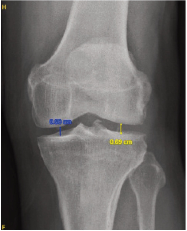
Results: Our study shows that reduction in joint space is more obvious in weight-bearing radiographs, however in severe or higher-grade osteoarthritis, a supine radiograph is adequate to diagnose knee osteoarthritis.
Conclusion: Standing radiograph of the knee is preferred to a supine knee radiograph wherever possible due to the additional value it brings, however, in certain patient conditions, a supine radiograph is still acceptable.
期刊介绍:
The Malaysian Orthopaedic Journal is a peer-reviewed journal that publishes original papers and case reports three times a year in both printed and electronic version. The purpose of MOJ is to disseminate new knowledge and provide updates in Orthopaedics, trauma and musculoskeletal research. It is an Open Access journal that does not require processing fee or article processing charge from the authors. The Malaysian Orthopaedic Journal is the official journal of Malaysian Orthopaedic Association (MOA) and ASEAN Orthopaedic Association (AOA).

 求助内容:
求助内容: 应助结果提醒方式:


