Edward A. FitzGerald, Daniela Cederfelt, Daria Kovryzhenko, Pierre Boronat, Bjarte Aarmo Lund, Doreen Dobritzsch, Sven Hennig, Pablo Porragas Paseiro, Iwan J. P. de Esch and U. Helena Danielson
{"title":"利用生物传感器和x射线晶体学检测和表征配体诱导的乙酰胆碱结合蛋白构象变化。","authors":"Edward A. FitzGerald, Daniela Cederfelt, Daria Kovryzhenko, Pierre Boronat, Bjarte Aarmo Lund, Doreen Dobritzsch, Sven Hennig, Pablo Porragas Paseiro, Iwan J. P. de Esch and U. Helena Danielson","doi":"10.1039/D5CB00041F","DOIUrl":null,"url":null,"abstract":"<p >Analysis of ligand-induced structural changes in proteins is challenging due to the lack of experimental methods suited for detection and characterisation of both ligand binding and induced structural changes. We have explored biosensors with different detection principles to study interactions between ligands and acetylcholine binding proteins (AChBPs), soluble homologues of Cys-loop ligand gated ion channels (LGICs) that undergo similar structural changes as LGICs upon ligand binding. X-ray crystallography was used to identify binding sites and establish if the detected conformational changes involved small changes in loop C or major structural changes in the pentamer associated with ion channel opening. Experiments were initially focused on ligands exhibiting complex surface plasmon resonance (SPR) biosensor sensorgrams or detected by second harmonic generation (SHG) biosensor analysis. Surface acoustic wave (SAW) and SHG biosensors confirmed that complexities in SPR data were indeed due to ligand-induced conformational changes. Grating coupled interferometry (GCI) biosensor sensorgrams were less complex, despite similar detection principles. switchSENSE biosensor analysis revealed that ligands resulted in either a compaction or expansion of the protein structure. X-ray crystallography of the protein–ligand complexes was only successful for 7 out of 12 ligands, despite nM–μM affinities. Crystals were not obtained for the two compounds shown by SHG analysis to induce large structural changes, while electron densities were not seen in the structures for some ligands. The work presented herein shows that several biosensor technologies have a unique capability to detect and discriminate binding and ligand induced conformational changes in proteins, also when interactions are rapid, weak and structural changes are small. However, they are complementary and provide different information.</p>","PeriodicalId":40691,"journal":{"name":"RSC Chemical Biology","volume":" 10","pages":" 1625-1639"},"PeriodicalIF":3.1000,"publicationDate":"2025-08-13","publicationTypes":"Journal Article","fieldsOfStudy":null,"isOpenAccess":false,"openAccessPdf":"https://www.ncbi.nlm.nih.gov/pmc/articles/PMC12394895/pdf/","citationCount":"0","resultStr":"{\"title\":\"Detection and characterisation of ligand-induced conformational changes in acetylcholine binding proteins using biosensors and X-ray crystallography\",\"authors\":\"Edward A. FitzGerald, Daniela Cederfelt, Daria Kovryzhenko, Pierre Boronat, Bjarte Aarmo Lund, Doreen Dobritzsch, Sven Hennig, Pablo Porragas Paseiro, Iwan J. P. de Esch and U. Helena Danielson\",\"doi\":\"10.1039/D5CB00041F\",\"DOIUrl\":null,\"url\":null,\"abstract\":\"<p >Analysis of ligand-induced structural changes in proteins is challenging due to the lack of experimental methods suited for detection and characterisation of both ligand binding and induced structural changes. We have explored biosensors with different detection principles to study interactions between ligands and acetylcholine binding proteins (AChBPs), soluble homologues of Cys-loop ligand gated ion channels (LGICs) that undergo similar structural changes as LGICs upon ligand binding. X-ray crystallography was used to identify binding sites and establish if the detected conformational changes involved small changes in loop C or major structural changes in the pentamer associated with ion channel opening. Experiments were initially focused on ligands exhibiting complex surface plasmon resonance (SPR) biosensor sensorgrams or detected by second harmonic generation (SHG) biosensor analysis. Surface acoustic wave (SAW) and SHG biosensors confirmed that complexities in SPR data were indeed due to ligand-induced conformational changes. Grating coupled interferometry (GCI) biosensor sensorgrams were less complex, despite similar detection principles. switchSENSE biosensor analysis revealed that ligands resulted in either a compaction or expansion of the protein structure. X-ray crystallography of the protein–ligand complexes was only successful for 7 out of 12 ligands, despite nM–μM affinities. Crystals were not obtained for the two compounds shown by SHG analysis to induce large structural changes, while electron densities were not seen in the structures for some ligands. The work presented herein shows that several biosensor technologies have a unique capability to detect and discriminate binding and ligand induced conformational changes in proteins, also when interactions are rapid, weak and structural changes are small. However, they are complementary and provide different information.</p>\",\"PeriodicalId\":40691,\"journal\":{\"name\":\"RSC Chemical Biology\",\"volume\":\" 10\",\"pages\":\" 1625-1639\"},\"PeriodicalIF\":3.1000,\"publicationDate\":\"2025-08-13\",\"publicationTypes\":\"Journal Article\",\"fieldsOfStudy\":null,\"isOpenAccess\":false,\"openAccessPdf\":\"https://www.ncbi.nlm.nih.gov/pmc/articles/PMC12394895/pdf/\",\"citationCount\":\"0\",\"resultStr\":null,\"platform\":\"Semanticscholar\",\"paperid\":null,\"PeriodicalName\":\"RSC Chemical Biology\",\"FirstCategoryId\":\"1085\",\"ListUrlMain\":\"https://pubs.rsc.org/en/content/articlelanding/2025/cb/d5cb00041f\",\"RegionNum\":0,\"RegionCategory\":null,\"ArticlePicture\":[],\"TitleCN\":null,\"AbstractTextCN\":null,\"PMCID\":null,\"EPubDate\":\"\",\"PubModel\":\"\",\"JCR\":\"Q2\",\"JCRName\":\"BIOCHEMISTRY & MOLECULAR BIOLOGY\",\"Score\":null,\"Total\":0}","platform":"Semanticscholar","paperid":null,"PeriodicalName":"RSC Chemical Biology","FirstCategoryId":"1085","ListUrlMain":"https://pubs.rsc.org/en/content/articlelanding/2025/cb/d5cb00041f","RegionNum":0,"RegionCategory":null,"ArticlePicture":[],"TitleCN":null,"AbstractTextCN":null,"PMCID":null,"EPubDate":"","PubModel":"","JCR":"Q2","JCRName":"BIOCHEMISTRY & MOLECULAR BIOLOGY","Score":null,"Total":0}
Detection and characterisation of ligand-induced conformational changes in acetylcholine binding proteins using biosensors and X-ray crystallography
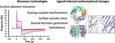
Analysis of ligand-induced structural changes in proteins is challenging due to the lack of experimental methods suited for detection and characterisation of both ligand binding and induced structural changes. We have explored biosensors with different detection principles to study interactions between ligands and acetylcholine binding proteins (AChBPs), soluble homologues of Cys-loop ligand gated ion channels (LGICs) that undergo similar structural changes as LGICs upon ligand binding. X-ray crystallography was used to identify binding sites and establish if the detected conformational changes involved small changes in loop C or major structural changes in the pentamer associated with ion channel opening. Experiments were initially focused on ligands exhibiting complex surface plasmon resonance (SPR) biosensor sensorgrams or detected by second harmonic generation (SHG) biosensor analysis. Surface acoustic wave (SAW) and SHG biosensors confirmed that complexities in SPR data were indeed due to ligand-induced conformational changes. Grating coupled interferometry (GCI) biosensor sensorgrams were less complex, despite similar detection principles. switchSENSE biosensor analysis revealed that ligands resulted in either a compaction or expansion of the protein structure. X-ray crystallography of the protein–ligand complexes was only successful for 7 out of 12 ligands, despite nM–μM affinities. Crystals were not obtained for the two compounds shown by SHG analysis to induce large structural changes, while electron densities were not seen in the structures for some ligands. The work presented herein shows that several biosensor technologies have a unique capability to detect and discriminate binding and ligand induced conformational changes in proteins, also when interactions are rapid, weak and structural changes are small. However, they are complementary and provide different information.

 求助内容:
求助内容: 应助结果提醒方式:


