T Tachibana, H Katagiri, T Ogawa, K Miyatake, R Takada, T Jinno
{"title":"基于三维(3D)分析的髋关节发育不良的股骨近端形态。","authors":"T Tachibana, H Katagiri, T Ogawa, K Miyatake, R Takada, T Jinno","doi":"10.5704/MOJ.2507.014","DOIUrl":null,"url":null,"abstract":"<p><strong>Introduction: </strong>Surgeons performing periacetabular osteotomy (PAO) should account for proximal femoral morphology to prevent secondary femoroacetabular impingement. Herein, we aimed to clarify proximal femoral morphology in patients with developmental dysplasia of the hip (DDH).</p><p><strong>Materials and methods: </strong>This retrospective study included 57 patients with DDH (77 hips) who underwent PAO (DDH group). The control group comprised 30 patients (30 hips) with unilateral femoral head necrosis and contralateral unaffected hips (healthy hips). Coronal planes were created parallel to the femoral neck axis based on three-dimensional image analysis of hip computed tomography images. Coronal slices were obtained using clockwise rotation around the femoral neck axis in 15° increments, creating seven positions for measuring alpha (α)-angles. The superior and anterior directions were defined as 12 o'clock and 3 o'clock, respectively. Cam deformity was defined as an α-angle ≥60°. Outcome measurements were the α-angles of seven slices, cam deformity, and correlations between the maximum value of the α-angles and related factors.</p><p><strong>Results: </strong>α-Angles were greater in the superior direction in the control than in the DDH group; conversely, they were greater in the anterior direction in the DDH than in the control group. The DDH group had more cam deformities than the control group. Cam deformities were more superior (12:30 to 1:00) in the control group, and more anterior (2:00 to 3:00) in the DDH group. Maximum α-angles in the DDH group correlated with superior acetabular coverage.</p><p><strong>Conclusion: </strong>Surgeons should carefully consider acetabular version during PAO and avoid acetabular retroversion in cases with cam deformities.</p>","PeriodicalId":45241,"journal":{"name":"Malaysian Orthopaedic Journal","volume":"19 2","pages":"108-116"},"PeriodicalIF":0.6000,"publicationDate":"2025-07-01","publicationTypes":"Journal Article","fieldsOfStudy":null,"isOpenAccess":false,"openAccessPdf":"https://www.ncbi.nlm.nih.gov/pmc/articles/PMC12368459/pdf/","citationCount":"0","resultStr":"{\"title\":\"Proximal Femoral Morphology in Development Dysplasia of the Hip Based on Three-Dimensional (3D) Analysis.\",\"authors\":\"T Tachibana, H Katagiri, T Ogawa, K Miyatake, R Takada, T Jinno\",\"doi\":\"10.5704/MOJ.2507.014\",\"DOIUrl\":null,\"url\":null,\"abstract\":\"<p><strong>Introduction: </strong>Surgeons performing periacetabular osteotomy (PAO) should account for proximal femoral morphology to prevent secondary femoroacetabular impingement. Herein, we aimed to clarify proximal femoral morphology in patients with developmental dysplasia of the hip (DDH).</p><p><strong>Materials and methods: </strong>This retrospective study included 57 patients with DDH (77 hips) who underwent PAO (DDH group). The control group comprised 30 patients (30 hips) with unilateral femoral head necrosis and contralateral unaffected hips (healthy hips). Coronal planes were created parallel to the femoral neck axis based on three-dimensional image analysis of hip computed tomography images. Coronal slices were obtained using clockwise rotation around the femoral neck axis in 15° increments, creating seven positions for measuring alpha (α)-angles. The superior and anterior directions were defined as 12 o'clock and 3 o'clock, respectively. Cam deformity was defined as an α-angle ≥60°. Outcome measurements were the α-angles of seven slices, cam deformity, and correlations between the maximum value of the α-angles and related factors.</p><p><strong>Results: </strong>α-Angles were greater in the superior direction in the control than in the DDH group; conversely, they were greater in the anterior direction in the DDH than in the control group. The DDH group had more cam deformities than the control group. Cam deformities were more superior (12:30 to 1:00) in the control group, and more anterior (2:00 to 3:00) in the DDH group. Maximum α-angles in the DDH group correlated with superior acetabular coverage.</p><p><strong>Conclusion: </strong>Surgeons should carefully consider acetabular version during PAO and avoid acetabular retroversion in cases with cam deformities.</p>\",\"PeriodicalId\":45241,\"journal\":{\"name\":\"Malaysian Orthopaedic Journal\",\"volume\":\"19 2\",\"pages\":\"108-116\"},\"PeriodicalIF\":0.6000,\"publicationDate\":\"2025-07-01\",\"publicationTypes\":\"Journal Article\",\"fieldsOfStudy\":null,\"isOpenAccess\":false,\"openAccessPdf\":\"https://www.ncbi.nlm.nih.gov/pmc/articles/PMC12368459/pdf/\",\"citationCount\":\"0\",\"resultStr\":null,\"platform\":\"Semanticscholar\",\"paperid\":null,\"PeriodicalName\":\"Malaysian Orthopaedic Journal\",\"FirstCategoryId\":\"1085\",\"ListUrlMain\":\"https://doi.org/10.5704/MOJ.2507.014\",\"RegionNum\":0,\"RegionCategory\":null,\"ArticlePicture\":[],\"TitleCN\":null,\"AbstractTextCN\":null,\"PMCID\":null,\"EPubDate\":\"\",\"PubModel\":\"\",\"JCR\":\"Q4\",\"JCRName\":\"ORTHOPEDICS\",\"Score\":null,\"Total\":0}","platform":"Semanticscholar","paperid":null,"PeriodicalName":"Malaysian Orthopaedic Journal","FirstCategoryId":"1085","ListUrlMain":"https://doi.org/10.5704/MOJ.2507.014","RegionNum":0,"RegionCategory":null,"ArticlePicture":[],"TitleCN":null,"AbstractTextCN":null,"PMCID":null,"EPubDate":"","PubModel":"","JCR":"Q4","JCRName":"ORTHOPEDICS","Score":null,"Total":0}
Proximal Femoral Morphology in Development Dysplasia of the Hip Based on Three-Dimensional (3D) Analysis.
Introduction: Surgeons performing periacetabular osteotomy (PAO) should account for proximal femoral morphology to prevent secondary femoroacetabular impingement. Herein, we aimed to clarify proximal femoral morphology in patients with developmental dysplasia of the hip (DDH).
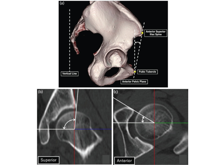
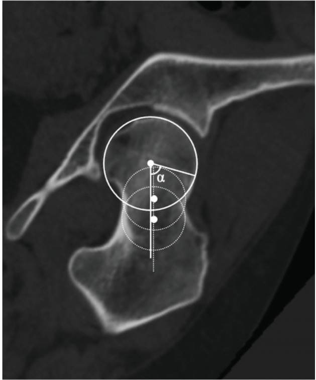
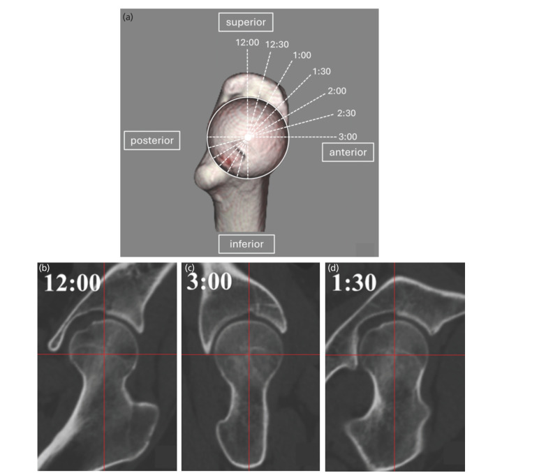
Materials and methods: This retrospective study included 57 patients with DDH (77 hips) who underwent PAO (DDH group). The control group comprised 30 patients (30 hips) with unilateral femoral head necrosis and contralateral unaffected hips (healthy hips). Coronal planes were created parallel to the femoral neck axis based on three-dimensional image analysis of hip computed tomography images. Coronal slices were obtained using clockwise rotation around the femoral neck axis in 15° increments, creating seven positions for measuring alpha (α)-angles. The superior and anterior directions were defined as 12 o'clock and 3 o'clock, respectively. Cam deformity was defined as an α-angle ≥60°. Outcome measurements were the α-angles of seven slices, cam deformity, and correlations between the maximum value of the α-angles and related factors.
Results: α-Angles were greater in the superior direction in the control than in the DDH group; conversely, they were greater in the anterior direction in the DDH than in the control group. The DDH group had more cam deformities than the control group. Cam deformities were more superior (12:30 to 1:00) in the control group, and more anterior (2:00 to 3:00) in the DDH group. Maximum α-angles in the DDH group correlated with superior acetabular coverage.
Conclusion: Surgeons should carefully consider acetabular version during PAO and avoid acetabular retroversion in cases with cam deformities.
期刊介绍:
The Malaysian Orthopaedic Journal is a peer-reviewed journal that publishes original papers and case reports three times a year in both printed and electronic version. The purpose of MOJ is to disseminate new knowledge and provide updates in Orthopaedics, trauma and musculoskeletal research. It is an Open Access journal that does not require processing fee or article processing charge from the authors. The Malaysian Orthopaedic Journal is the official journal of Malaysian Orthopaedic Association (MOA) and ASEAN Orthopaedic Association (AOA).

 求助内容:
求助内容: 应助结果提醒方式:


