Nicole Chan, Ronald P Kaufman, Nada Farhat, Grace Y Kim
{"title":"嗜铬细胞瘤1例,生化检查阳性,无肿瘤。","authors":"Nicole Chan, Ronald P Kaufman, Nada Farhat, Grace Y Kim","doi":"10.1530/EDM-25-0027","DOIUrl":null,"url":null,"abstract":"<p><strong>Summary: </strong>Pheochromocytomas are rare neuroendocrine tumors derived from adrenal chromaffin cells that result in hyperactivity of the sympathetic nervous system. We present the case of a patient with biochemical evidence of pheochromocytoma, but surgical pathology revealed absence of tumor. This is an 80-year-old female with a past medical history of metastatic follicular lymphoma and hypertension with an incidental 1.4 cm right-sided adrenal nodule noted on a PET-CT scan. Her hypertension was treated with three different antihypertensive agents. Subsequent imaging with non-contrast CT of abdomen and pelvis showed a right adrenal incidentaloma with 16 Hounsfield units. Abdominal MRI with and without contrast revealed atypical signal loss on the out-of-phase imaging. Plasma normetanephrines were approximately 2.4 times higher than the upper limit of normal. Her urinary normetanephrines were higher than the upper limit of normal, suggestive of pheochromocytoma. The patient proceeded with robotically assisted laparoscopic right adrenalectomy and postoperatively required vasopressors. Surgical pathology showed adrenal cortical hyperplasia and medullary infarction associated with fibrosis. However, the noted phases of necrosis with predominant fibrosis match the time interval between clinical diagnosis and surgical management. Postoperative metanephrines normalized 4 weeks after surgery, indicating successful surgical resection of autonomous secretion of metanephrines. This is the only known case of biochemical evidence of pheochromocytoma with no histologic evidence of tumor.</p><p><strong>Learning points: </strong>Biochemical evidence, clinical presentation and imaging studies of pheochromocytoma are crucial for its diagnosis. Due to the vascular nature of pheochromocytoma, there is a potential for the tumor to infarct. Plasma normetanephrines of greater than twice the upper limit of normal have high specificity for pheochromocytoma.</p>","PeriodicalId":37467,"journal":{"name":"Endocrinology, Diabetes and Metabolism Case Reports","volume":"2025 3","pages":""},"PeriodicalIF":0.7000,"publicationDate":"2025-09-02","publicationTypes":"Journal Article","fieldsOfStudy":null,"isOpenAccess":false,"openAccessPdf":"https://www.ncbi.nlm.nih.gov/pmc/articles/PMC12412360/pdf/","citationCount":"0","resultStr":"{\"title\":\"An uncommon case of pheochromocytoma with positive biochemical workup and absence of tumor.\",\"authors\":\"Nicole Chan, Ronald P Kaufman, Nada Farhat, Grace Y Kim\",\"doi\":\"10.1530/EDM-25-0027\",\"DOIUrl\":null,\"url\":null,\"abstract\":\"<p><strong>Summary: </strong>Pheochromocytomas are rare neuroendocrine tumors derived from adrenal chromaffin cells that result in hyperactivity of the sympathetic nervous system. We present the case of a patient with biochemical evidence of pheochromocytoma, but surgical pathology revealed absence of tumor. This is an 80-year-old female with a past medical history of metastatic follicular lymphoma and hypertension with an incidental 1.4 cm right-sided adrenal nodule noted on a PET-CT scan. Her hypertension was treated with three different antihypertensive agents. Subsequent imaging with non-contrast CT of abdomen and pelvis showed a right adrenal incidentaloma with 16 Hounsfield units. Abdominal MRI with and without contrast revealed atypical signal loss on the out-of-phase imaging. Plasma normetanephrines were approximately 2.4 times higher than the upper limit of normal. Her urinary normetanephrines were higher than the upper limit of normal, suggestive of pheochromocytoma. The patient proceeded with robotically assisted laparoscopic right adrenalectomy and postoperatively required vasopressors. Surgical pathology showed adrenal cortical hyperplasia and medullary infarction associated with fibrosis. However, the noted phases of necrosis with predominant fibrosis match the time interval between clinical diagnosis and surgical management. Postoperative metanephrines normalized 4 weeks after surgery, indicating successful surgical resection of autonomous secretion of metanephrines. This is the only known case of biochemical evidence of pheochromocytoma with no histologic evidence of tumor.</p><p><strong>Learning points: </strong>Biochemical evidence, clinical presentation and imaging studies of pheochromocytoma are crucial for its diagnosis. Due to the vascular nature of pheochromocytoma, there is a potential for the tumor to infarct. Plasma normetanephrines of greater than twice the upper limit of normal have high specificity for pheochromocytoma.</p>\",\"PeriodicalId\":37467,\"journal\":{\"name\":\"Endocrinology, Diabetes and Metabolism Case Reports\",\"volume\":\"2025 3\",\"pages\":\"\"},\"PeriodicalIF\":0.7000,\"publicationDate\":\"2025-09-02\",\"publicationTypes\":\"Journal Article\",\"fieldsOfStudy\":null,\"isOpenAccess\":false,\"openAccessPdf\":\"https://www.ncbi.nlm.nih.gov/pmc/articles/PMC12412360/pdf/\",\"citationCount\":\"0\",\"resultStr\":null,\"platform\":\"Semanticscholar\",\"paperid\":null,\"PeriodicalName\":\"Endocrinology, Diabetes and Metabolism Case Reports\",\"FirstCategoryId\":\"1085\",\"ListUrlMain\":\"https://doi.org/10.1530/EDM-25-0027\",\"RegionNum\":0,\"RegionCategory\":null,\"ArticlePicture\":[],\"TitleCN\":null,\"AbstractTextCN\":null,\"PMCID\":null,\"EPubDate\":\"2025/7/1 0:00:00\",\"PubModel\":\"Print\",\"JCR\":\"Q4\",\"JCRName\":\"ENDOCRINOLOGY & METABOLISM\",\"Score\":null,\"Total\":0}","platform":"Semanticscholar","paperid":null,"PeriodicalName":"Endocrinology, Diabetes and Metabolism Case Reports","FirstCategoryId":"1085","ListUrlMain":"https://doi.org/10.1530/EDM-25-0027","RegionNum":0,"RegionCategory":null,"ArticlePicture":[],"TitleCN":null,"AbstractTextCN":null,"PMCID":null,"EPubDate":"2025/7/1 0:00:00","PubModel":"Print","JCR":"Q4","JCRName":"ENDOCRINOLOGY & METABOLISM","Score":null,"Total":0}
An uncommon case of pheochromocytoma with positive biochemical workup and absence of tumor.
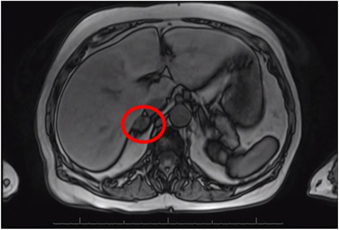
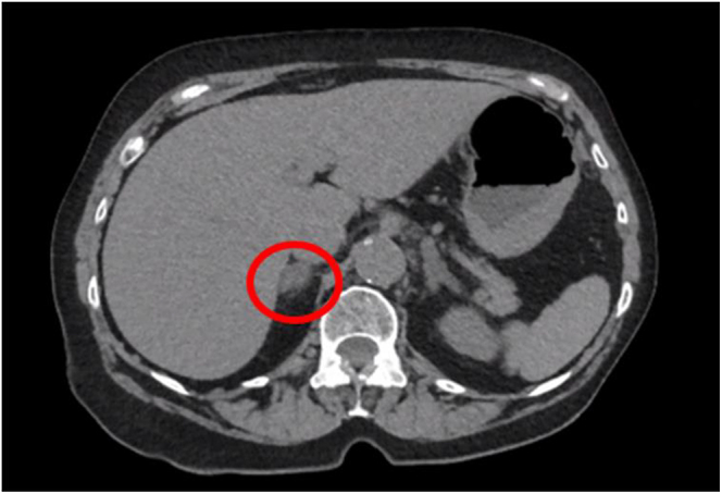
Summary: Pheochromocytomas are rare neuroendocrine tumors derived from adrenal chromaffin cells that result in hyperactivity of the sympathetic nervous system. We present the case of a patient with biochemical evidence of pheochromocytoma, but surgical pathology revealed absence of tumor. This is an 80-year-old female with a past medical history of metastatic follicular lymphoma and hypertension with an incidental 1.4 cm right-sided adrenal nodule noted on a PET-CT scan. Her hypertension was treated with three different antihypertensive agents. Subsequent imaging with non-contrast CT of abdomen and pelvis showed a right adrenal incidentaloma with 16 Hounsfield units. Abdominal MRI with and without contrast revealed atypical signal loss on the out-of-phase imaging. Plasma normetanephrines were approximately 2.4 times higher than the upper limit of normal. Her urinary normetanephrines were higher than the upper limit of normal, suggestive of pheochromocytoma. The patient proceeded with robotically assisted laparoscopic right adrenalectomy and postoperatively required vasopressors. Surgical pathology showed adrenal cortical hyperplasia and medullary infarction associated with fibrosis. However, the noted phases of necrosis with predominant fibrosis match the time interval between clinical diagnosis and surgical management. Postoperative metanephrines normalized 4 weeks after surgery, indicating successful surgical resection of autonomous secretion of metanephrines. This is the only known case of biochemical evidence of pheochromocytoma with no histologic evidence of tumor.
Learning points: Biochemical evidence, clinical presentation and imaging studies of pheochromocytoma are crucial for its diagnosis. Due to the vascular nature of pheochromocytoma, there is a potential for the tumor to infarct. Plasma normetanephrines of greater than twice the upper limit of normal have high specificity for pheochromocytoma.
期刊介绍:
Endocrinology, Diabetes & Metabolism Case Reports publishes case reports on common and rare conditions in all areas of clinical endocrinology, diabetes and metabolism. Articles should include clear learning points which readers can use to inform medical education or clinical practice. The types of cases of interest to Endocrinology, Diabetes & Metabolism Case Reports include: -Insight into disease pathogenesis or mechanism of therapy - Novel diagnostic procedure - Novel treatment - Unique/unexpected symptoms or presentations of a disease - New disease or syndrome: presentations/diagnosis/management - Unusual effects of medical treatment - Error in diagnosis/pitfalls and caveats


 求助内容:
求助内容: 应助结果提醒方式:


