{"title":"从检测到诊断:使用YOLO11和形态学后处理进行脑肿瘤MRI图像分析的高级迁移学习管道。","authors":"Ikram Chourib","doi":"10.3390/jimaging11080282","DOIUrl":null,"url":null,"abstract":"<p><p>Accurate and timely detection of brain tumors from magnetic resonance imaging (MRI) scans is critical for improving patient outcomes and informing therapeutic decision-making. However, the complex heterogeneity of tumor morphology, scarcity of annotated medical data, and computational demands of deep learning models present substantial challenges for developing reliable automated diagnostic systems. In this study, we propose a robust and scalable deep learning framework for brain tumor detection and classification, built upon an enhanced YOLO-v11 architecture combined with a two-stage transfer learning strategy. The first stage involves training a base model on a large, diverse MRI dataset. Upon achieving a mean Average Precision (mAP) exceeding 90%, this model is designated as the Brain Tumor Detection Model (BTDM). In the second stage, the BTDM is fine-tuned on a structurally similar but smaller dataset to form Brain Tumor Detection and Segmentation (BTDS), effectively leveraging domain transfer to maintain performance despite limited data. The model is further optimized through domain-specific data augmentation-including geometric transformations-to improve generalization and robustness. Experimental evaluations on publicly available datasets show that the framework achieves high mAP@0.5 scores (up to 93.5% for the BTDM and 91% for BTDS) and consistently outperforms existing state-of-the-art methods across multiple tumor types, including glioma, meningioma, and pituitary tumors. In addition, a post-processing module enhances interpretability by generating segmentation masks and extracting clinically relevant metrics such as tumor size and severity level. These results underscore the potential of our approach as a high-performance, interpretable, and deployable clinical decision-support tool, contributing to the advancement of intelligent real-time neuro-oncological diagnostics.</p>","PeriodicalId":37035,"journal":{"name":"Journal of Imaging","volume":"11 8","pages":""},"PeriodicalIF":2.7000,"publicationDate":"2025-08-21","publicationTypes":"Journal Article","fieldsOfStudy":null,"isOpenAccess":false,"openAccessPdf":"https://www.ncbi.nlm.nih.gov/pmc/articles/PMC12387851/pdf/","citationCount":"0","resultStr":"{\"title\":\"From Detection to Diagnosis: An Advanced Transfer Learning Pipeline Using YOLO11 with Morphological Post-Processing for Brain Tumor Analysis for MRI Images.\",\"authors\":\"Ikram Chourib\",\"doi\":\"10.3390/jimaging11080282\",\"DOIUrl\":null,\"url\":null,\"abstract\":\"<p><p>Accurate and timely detection of brain tumors from magnetic resonance imaging (MRI) scans is critical for improving patient outcomes and informing therapeutic decision-making. However, the complex heterogeneity of tumor morphology, scarcity of annotated medical data, and computational demands of deep learning models present substantial challenges for developing reliable automated diagnostic systems. In this study, we propose a robust and scalable deep learning framework for brain tumor detection and classification, built upon an enhanced YOLO-v11 architecture combined with a two-stage transfer learning strategy. The first stage involves training a base model on a large, diverse MRI dataset. Upon achieving a mean Average Precision (mAP) exceeding 90%, this model is designated as the Brain Tumor Detection Model (BTDM). In the second stage, the BTDM is fine-tuned on a structurally similar but smaller dataset to form Brain Tumor Detection and Segmentation (BTDS), effectively leveraging domain transfer to maintain performance despite limited data. The model is further optimized through domain-specific data augmentation-including geometric transformations-to improve generalization and robustness. Experimental evaluations on publicly available datasets show that the framework achieves high mAP@0.5 scores (up to 93.5% for the BTDM and 91% for BTDS) and consistently outperforms existing state-of-the-art methods across multiple tumor types, including glioma, meningioma, and pituitary tumors. In addition, a post-processing module enhances interpretability by generating segmentation masks and extracting clinically relevant metrics such as tumor size and severity level. These results underscore the potential of our approach as a high-performance, interpretable, and deployable clinical decision-support tool, contributing to the advancement of intelligent real-time neuro-oncological diagnostics.</p>\",\"PeriodicalId\":37035,\"journal\":{\"name\":\"Journal of Imaging\",\"volume\":\"11 8\",\"pages\":\"\"},\"PeriodicalIF\":2.7000,\"publicationDate\":\"2025-08-21\",\"publicationTypes\":\"Journal Article\",\"fieldsOfStudy\":null,\"isOpenAccess\":false,\"openAccessPdf\":\"https://www.ncbi.nlm.nih.gov/pmc/articles/PMC12387851/pdf/\",\"citationCount\":\"0\",\"resultStr\":null,\"platform\":\"Semanticscholar\",\"paperid\":null,\"PeriodicalName\":\"Journal of Imaging\",\"FirstCategoryId\":\"1085\",\"ListUrlMain\":\"https://doi.org/10.3390/jimaging11080282\",\"RegionNum\":0,\"RegionCategory\":null,\"ArticlePicture\":[],\"TitleCN\":null,\"AbstractTextCN\":null,\"PMCID\":null,\"EPubDate\":\"\",\"PubModel\":\"\",\"JCR\":\"Q3\",\"JCRName\":\"IMAGING SCIENCE & PHOTOGRAPHIC TECHNOLOGY\",\"Score\":null,\"Total\":0}","platform":"Semanticscholar","paperid":null,"PeriodicalName":"Journal of Imaging","FirstCategoryId":"1085","ListUrlMain":"https://doi.org/10.3390/jimaging11080282","RegionNum":0,"RegionCategory":null,"ArticlePicture":[],"TitleCN":null,"AbstractTextCN":null,"PMCID":null,"EPubDate":"","PubModel":"","JCR":"Q3","JCRName":"IMAGING SCIENCE & PHOTOGRAPHIC TECHNOLOGY","Score":null,"Total":0}
From Detection to Diagnosis: An Advanced Transfer Learning Pipeline Using YOLO11 with Morphological Post-Processing for Brain Tumor Analysis for MRI Images.
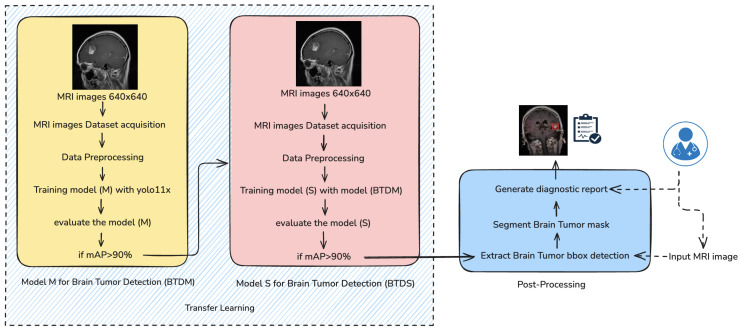
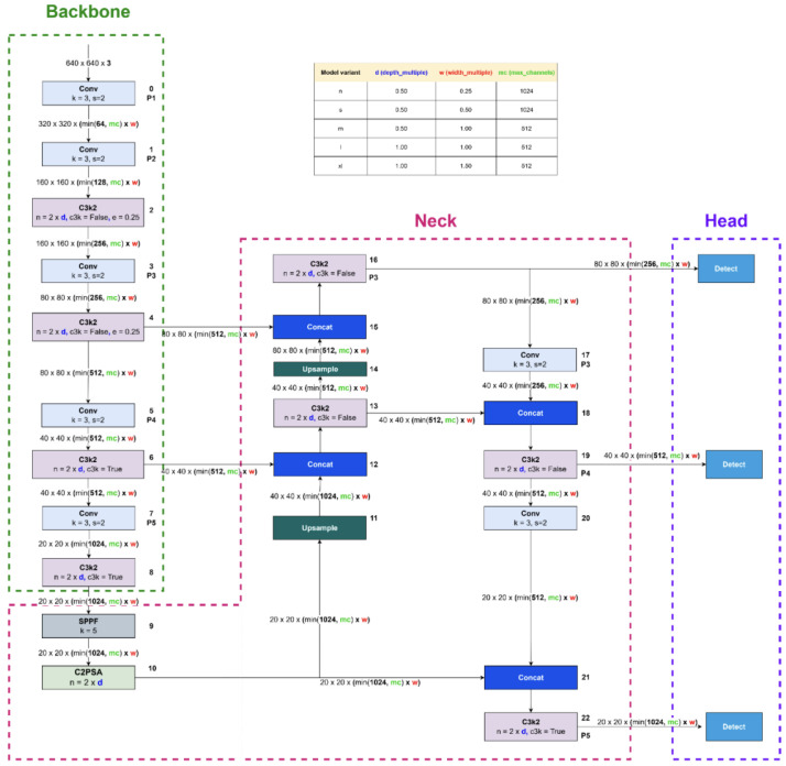
Accurate and timely detection of brain tumors from magnetic resonance imaging (MRI) scans is critical for improving patient outcomes and informing therapeutic decision-making. However, the complex heterogeneity of tumor morphology, scarcity of annotated medical data, and computational demands of deep learning models present substantial challenges for developing reliable automated diagnostic systems. In this study, we propose a robust and scalable deep learning framework for brain tumor detection and classification, built upon an enhanced YOLO-v11 architecture combined with a two-stage transfer learning strategy. The first stage involves training a base model on a large, diverse MRI dataset. Upon achieving a mean Average Precision (mAP) exceeding 90%, this model is designated as the Brain Tumor Detection Model (BTDM). In the second stage, the BTDM is fine-tuned on a structurally similar but smaller dataset to form Brain Tumor Detection and Segmentation (BTDS), effectively leveraging domain transfer to maintain performance despite limited data. The model is further optimized through domain-specific data augmentation-including geometric transformations-to improve generalization and robustness. Experimental evaluations on publicly available datasets show that the framework achieves high mAP@0.5 scores (up to 93.5% for the BTDM and 91% for BTDS) and consistently outperforms existing state-of-the-art methods across multiple tumor types, including glioma, meningioma, and pituitary tumors. In addition, a post-processing module enhances interpretability by generating segmentation masks and extracting clinically relevant metrics such as tumor size and severity level. These results underscore the potential of our approach as a high-performance, interpretable, and deployable clinical decision-support tool, contributing to the advancement of intelligent real-time neuro-oncological diagnostics.

 求助内容:
求助内容: 应助结果提醒方式:


