Cecilia Diana-Albelda, Álvaro García-Martín, Jesus Bescos
{"title":"神经胶质瘤分割的深度学习方法综述,局限性和未来展望。","authors":"Cecilia Diana-Albelda, Álvaro García-Martín, Jesus Bescos","doi":"10.3390/jimaging11080269","DOIUrl":null,"url":null,"abstract":"<p><p>Accurate and automated segmentation of gliomas from Magnetic Resonance Imaging (MRI) is crucial for effective diagnosis, treatment planning, and patient monitoring. However, the aggressive nature and morphological complexity of these tumors pose significant challenges that call for advanced segmentation techniques. This review provides a comprehensive analysis of Deep Learning (DL) methods for glioma segmentation, with a specific focus on bridging the gap between research performance and practical clinical deployment. We evaluate over 80 state-of-the-art models published up to 2025, categorizing them into CNN-based, Pure Transformer, and Hybrid CNN-Transformer architectures. The primary objective of this paper is to critically assess these models not only on their segmentation accuracy but also on their computational efficiency and suitability for real-world medical environments by incorporating hardware resource considerations. We present a comparison of model performance on the BraTS datasets benchmark and introduce a suitability analysis for top-performing models based on their robustness, efficiency, and completeness of tumor region delineation. By identifying current trends, limitations, and key trade-offs, this review offers future research directions aimed at optimizing the balance between technical performance and clinical usability to improve diagnostic outcomes for glioma patients.</p>","PeriodicalId":37035,"journal":{"name":"Journal of Imaging","volume":"11 8","pages":""},"PeriodicalIF":2.7000,"publicationDate":"2025-08-11","publicationTypes":"Journal Article","fieldsOfStudy":null,"isOpenAccess":false,"openAccessPdf":"https://www.ncbi.nlm.nih.gov/pmc/articles/PMC12387613/pdf/","citationCount":"0","resultStr":"{\"title\":\"A Review on Deep Learning Methods for Glioma Segmentation, Limitations, and Future Perspectives.\",\"authors\":\"Cecilia Diana-Albelda, Álvaro García-Martín, Jesus Bescos\",\"doi\":\"10.3390/jimaging11080269\",\"DOIUrl\":null,\"url\":null,\"abstract\":\"<p><p>Accurate and automated segmentation of gliomas from Magnetic Resonance Imaging (MRI) is crucial for effective diagnosis, treatment planning, and patient monitoring. However, the aggressive nature and morphological complexity of these tumors pose significant challenges that call for advanced segmentation techniques. This review provides a comprehensive analysis of Deep Learning (DL) methods for glioma segmentation, with a specific focus on bridging the gap between research performance and practical clinical deployment. We evaluate over 80 state-of-the-art models published up to 2025, categorizing them into CNN-based, Pure Transformer, and Hybrid CNN-Transformer architectures. The primary objective of this paper is to critically assess these models not only on their segmentation accuracy but also on their computational efficiency and suitability for real-world medical environments by incorporating hardware resource considerations. We present a comparison of model performance on the BraTS datasets benchmark and introduce a suitability analysis for top-performing models based on their robustness, efficiency, and completeness of tumor region delineation. By identifying current trends, limitations, and key trade-offs, this review offers future research directions aimed at optimizing the balance between technical performance and clinical usability to improve diagnostic outcomes for glioma patients.</p>\",\"PeriodicalId\":37035,\"journal\":{\"name\":\"Journal of Imaging\",\"volume\":\"11 8\",\"pages\":\"\"},\"PeriodicalIF\":2.7000,\"publicationDate\":\"2025-08-11\",\"publicationTypes\":\"Journal Article\",\"fieldsOfStudy\":null,\"isOpenAccess\":false,\"openAccessPdf\":\"https://www.ncbi.nlm.nih.gov/pmc/articles/PMC12387613/pdf/\",\"citationCount\":\"0\",\"resultStr\":null,\"platform\":\"Semanticscholar\",\"paperid\":null,\"PeriodicalName\":\"Journal of Imaging\",\"FirstCategoryId\":\"1085\",\"ListUrlMain\":\"https://doi.org/10.3390/jimaging11080269\",\"RegionNum\":0,\"RegionCategory\":null,\"ArticlePicture\":[],\"TitleCN\":null,\"AbstractTextCN\":null,\"PMCID\":null,\"EPubDate\":\"\",\"PubModel\":\"\",\"JCR\":\"Q3\",\"JCRName\":\"IMAGING SCIENCE & PHOTOGRAPHIC TECHNOLOGY\",\"Score\":null,\"Total\":0}","platform":"Semanticscholar","paperid":null,"PeriodicalName":"Journal of Imaging","FirstCategoryId":"1085","ListUrlMain":"https://doi.org/10.3390/jimaging11080269","RegionNum":0,"RegionCategory":null,"ArticlePicture":[],"TitleCN":null,"AbstractTextCN":null,"PMCID":null,"EPubDate":"","PubModel":"","JCR":"Q3","JCRName":"IMAGING SCIENCE & PHOTOGRAPHIC TECHNOLOGY","Score":null,"Total":0}
A Review on Deep Learning Methods for Glioma Segmentation, Limitations, and Future Perspectives.
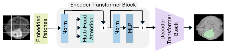
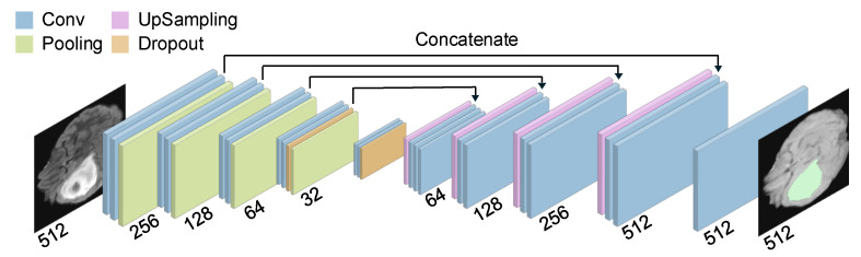
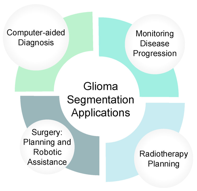
Accurate and automated segmentation of gliomas from Magnetic Resonance Imaging (MRI) is crucial for effective diagnosis, treatment planning, and patient monitoring. However, the aggressive nature and morphological complexity of these tumors pose significant challenges that call for advanced segmentation techniques. This review provides a comprehensive analysis of Deep Learning (DL) methods for glioma segmentation, with a specific focus on bridging the gap between research performance and practical clinical deployment. We evaluate over 80 state-of-the-art models published up to 2025, categorizing them into CNN-based, Pure Transformer, and Hybrid CNN-Transformer architectures. The primary objective of this paper is to critically assess these models not only on their segmentation accuracy but also on their computational efficiency and suitability for real-world medical environments by incorporating hardware resource considerations. We present a comparison of model performance on the BraTS datasets benchmark and introduce a suitability analysis for top-performing models based on their robustness, efficiency, and completeness of tumor region delineation. By identifying current trends, limitations, and key trade-offs, this review offers future research directions aimed at optimizing the balance between technical performance and clinical usability to improve diagnostic outcomes for glioma patients.

 求助内容:
求助内容: 应助结果提醒方式:


