Zhuoqun Zheng, Jin Ding, Yanping Chen, Hongjun Hua
{"title":"十二指肠乳头状病变内镜钳活检与切除标本的组织病理学差异:回顾性研究。","authors":"Zhuoqun Zheng, Jin Ding, Yanping Chen, Hongjun Hua","doi":"10.1186/s12876-025-04255-6","DOIUrl":null,"url":null,"abstract":"<p><strong>Background: </strong>Endoscopic papillectomy (EP) has been used for the treatment of duodenal papillectomy, and the results of preoperative endoscopic biopsy are important for the selection of treatment plans for the patients. However, some lesions cannot be precisely diagnosed based solely on biopsy results. In the study, we attempted to assess the accuracy of histopathological diagnosis of endoscopic forceps biopsy and to avoid over - or under-treatment.</p><p><strong>Methods: </strong>This retrospective observational analysis involved clinical data and endoscopic images of lesions diagnosed as non-cancerous on preoperative biopsy at the Gastroenterology Department of Jinhua Central Hospital from January 2021 to December 2024 and received follow-up treatment. We assessed the histological discrepancies between endoscopic forceps biopsy and specimens, and analyzed their correlation with clinical characteristics.</p><p><strong>Results: </strong>The study included 58 patients. The discrepancy rate between forceps biopsy and resected specimens was 50% (29/58 ). 28 of the 29 inconsistent biopsies were diagnosed with a higher grade, and the other one was diagnosed as a neuroendocrine tumor. 8 of the 40 biopsies that were diagnosed as indefinite for neoplasia or low-grade intraepithelial neoplasia were upgraded to HGIN or ampullary cancer after resection, which was related to duct (pancreatic duct or bile duct) dilatation (p = 0.003). Moreover, 13 of the 18 biopsies that were diagnosed with HGIN were upgraded to ampullary cancer after resection, which was related to lesion size (p = 0.035), and gender (p = 0.008). According to the postoperative pathological results, the lesions were divided into benign lesions and the malignant(HGIN and ampullary cancer), and histological discrepancy was associated with lesion size (p = 0.008), color (p = 0.000), and duct dilatation ( p = 0.001). Logistic regression analysis revealed that lesion size(OR = 3.566, 95%CI:1.085 ~ 11.723, P = 0.036) was a significant predictor of malignancy in ampullary adenomas.</p><p><strong>Conclusions: </strong>Histological discrepancies between endoscopic forceps biopsy and resected specimens of ampullary lesions were commonly observed in clinical practice. The presence of symptoms, including red color, lesion size > 1.25 cm and duct dilatation on radiologic imaging suggests the possibility of malignancy in ampullary adenoma.</p>","PeriodicalId":9129,"journal":{"name":"BMC Gastroenterology","volume":"25 1","pages":"632"},"PeriodicalIF":2.5000,"publicationDate":"2025-09-02","publicationTypes":"Journal Article","fieldsOfStudy":null,"isOpenAccess":false,"openAccessPdf":"https://www.ncbi.nlm.nih.gov/pmc/articles/PMC12403938/pdf/","citationCount":"0","resultStr":"{\"title\":\"Histopathologic discrepancies between endoscopic forceps biopsy and resection specimens in duodenal papillary lesions: a retrospective study.\",\"authors\":\"Zhuoqun Zheng, Jin Ding, Yanping Chen, Hongjun Hua\",\"doi\":\"10.1186/s12876-025-04255-6\",\"DOIUrl\":null,\"url\":null,\"abstract\":\"<p><strong>Background: </strong>Endoscopic papillectomy (EP) has been used for the treatment of duodenal papillectomy, and the results of preoperative endoscopic biopsy are important for the selection of treatment plans for the patients. However, some lesions cannot be precisely diagnosed based solely on biopsy results. In the study, we attempted to assess the accuracy of histopathological diagnosis of endoscopic forceps biopsy and to avoid over - or under-treatment.</p><p><strong>Methods: </strong>This retrospective observational analysis involved clinical data and endoscopic images of lesions diagnosed as non-cancerous on preoperative biopsy at the Gastroenterology Department of Jinhua Central Hospital from January 2021 to December 2024 and received follow-up treatment. We assessed the histological discrepancies between endoscopic forceps biopsy and specimens, and analyzed their correlation with clinical characteristics.</p><p><strong>Results: </strong>The study included 58 patients. The discrepancy rate between forceps biopsy and resected specimens was 50% (29/58 ). 28 of the 29 inconsistent biopsies were diagnosed with a higher grade, and the other one was diagnosed as a neuroendocrine tumor. 8 of the 40 biopsies that were diagnosed as indefinite for neoplasia or low-grade intraepithelial neoplasia were upgraded to HGIN or ampullary cancer after resection, which was related to duct (pancreatic duct or bile duct) dilatation (p = 0.003). Moreover, 13 of the 18 biopsies that were diagnosed with HGIN were upgraded to ampullary cancer after resection, which was related to lesion size (p = 0.035), and gender (p = 0.008). According to the postoperative pathological results, the lesions were divided into benign lesions and the malignant(HGIN and ampullary cancer), and histological discrepancy was associated with lesion size (p = 0.008), color (p = 0.000), and duct dilatation ( p = 0.001). Logistic regression analysis revealed that lesion size(OR = 3.566, 95%CI:1.085 ~ 11.723, P = 0.036) was a significant predictor of malignancy in ampullary adenomas.</p><p><strong>Conclusions: </strong>Histological discrepancies between endoscopic forceps biopsy and resected specimens of ampullary lesions were commonly observed in clinical practice. The presence of symptoms, including red color, lesion size > 1.25 cm and duct dilatation on radiologic imaging suggests the possibility of malignancy in ampullary adenoma.</p>\",\"PeriodicalId\":9129,\"journal\":{\"name\":\"BMC Gastroenterology\",\"volume\":\"25 1\",\"pages\":\"632\"},\"PeriodicalIF\":2.5000,\"publicationDate\":\"2025-09-02\",\"publicationTypes\":\"Journal Article\",\"fieldsOfStudy\":null,\"isOpenAccess\":false,\"openAccessPdf\":\"https://www.ncbi.nlm.nih.gov/pmc/articles/PMC12403938/pdf/\",\"citationCount\":\"0\",\"resultStr\":null,\"platform\":\"Semanticscholar\",\"paperid\":null,\"PeriodicalName\":\"BMC Gastroenterology\",\"FirstCategoryId\":\"3\",\"ListUrlMain\":\"https://doi.org/10.1186/s12876-025-04255-6\",\"RegionNum\":3,\"RegionCategory\":\"医学\",\"ArticlePicture\":[],\"TitleCN\":null,\"AbstractTextCN\":null,\"PMCID\":null,\"EPubDate\":\"\",\"PubModel\":\"\",\"JCR\":\"Q2\",\"JCRName\":\"GASTROENTEROLOGY & HEPATOLOGY\",\"Score\":null,\"Total\":0}","platform":"Semanticscholar","paperid":null,"PeriodicalName":"BMC Gastroenterology","FirstCategoryId":"3","ListUrlMain":"https://doi.org/10.1186/s12876-025-04255-6","RegionNum":3,"RegionCategory":"医学","ArticlePicture":[],"TitleCN":null,"AbstractTextCN":null,"PMCID":null,"EPubDate":"","PubModel":"","JCR":"Q2","JCRName":"GASTROENTEROLOGY & HEPATOLOGY","Score":null,"Total":0}
Histopathologic discrepancies between endoscopic forceps biopsy and resection specimens in duodenal papillary lesions: a retrospective study.
Background: Endoscopic papillectomy (EP) has been used for the treatment of duodenal papillectomy, and the results of preoperative endoscopic biopsy are important for the selection of treatment plans for the patients. However, some lesions cannot be precisely diagnosed based solely on biopsy results. In the study, we attempted to assess the accuracy of histopathological diagnosis of endoscopic forceps biopsy and to avoid over - or under-treatment.
Methods: This retrospective observational analysis involved clinical data and endoscopic images of lesions diagnosed as non-cancerous on preoperative biopsy at the Gastroenterology Department of Jinhua Central Hospital from January 2021 to December 2024 and received follow-up treatment. We assessed the histological discrepancies between endoscopic forceps biopsy and specimens, and analyzed their correlation with clinical characteristics.
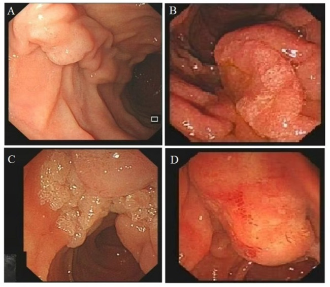
Results: The study included 58 patients. The discrepancy rate between forceps biopsy and resected specimens was 50% (29/58 ). 28 of the 29 inconsistent biopsies were diagnosed with a higher grade, and the other one was diagnosed as a neuroendocrine tumor. 8 of the 40 biopsies that were diagnosed as indefinite for neoplasia or low-grade intraepithelial neoplasia were upgraded to HGIN or ampullary cancer after resection, which was related to duct (pancreatic duct or bile duct) dilatation (p = 0.003). Moreover, 13 of the 18 biopsies that were diagnosed with HGIN were upgraded to ampullary cancer after resection, which was related to lesion size (p = 0.035), and gender (p = 0.008). According to the postoperative pathological results, the lesions were divided into benign lesions and the malignant(HGIN and ampullary cancer), and histological discrepancy was associated with lesion size (p = 0.008), color (p = 0.000), and duct dilatation ( p = 0.001). Logistic regression analysis revealed that lesion size(OR = 3.566, 95%CI:1.085 ~ 11.723, P = 0.036) was a significant predictor of malignancy in ampullary adenomas.
Conclusions: Histological discrepancies between endoscopic forceps biopsy and resected specimens of ampullary lesions were commonly observed in clinical practice. The presence of symptoms, including red color, lesion size > 1.25 cm and duct dilatation on radiologic imaging suggests the possibility of malignancy in ampullary adenoma.
期刊介绍:
BMC Gastroenterology is an open access, peer-reviewed journal that considers articles on all aspects of the prevention, diagnosis and management of gastrointestinal and hepatobiliary disorders, as well as related molecular genetics, pathophysiology, and epidemiology.
 求助内容:
求助内容: 应助结果提醒方式:


