{"title":"基于背景身体信号抑制的弥散加权全身成像血管炎症可视化。","authors":"Ayaka Ohno, Kenjuro Higo, Sawako Hiwatari, Takeko Kawabata, Hitoshi Nakashima, Mitsuru Ohishi","doi":"10.3400/avd.cr.25-00030","DOIUrl":null,"url":null,"abstract":"<p><p>Diffusion-weighted whole-body imaging with background body signal suppression has been used to diagnose fever of unknown origin. An 86-year-old man who underwent bile duct jejunostomy for bile duct cancer presented with fever (body temperature, 40°C). <i>Escherichia coli</i> was detected in blood cultures. Diffusion-weighted whole-body imaging with background body signal suppression revealed accumulation in the aortic arch. Therefore, infectious aortitis secondary to retrograde cholangitis was diagnosed. The patient was treated with antibiotics, and the aortic arch accumulation disappeared. Diffusion-weighted whole-body imaging with background body signal suppression is a useful modality for diagnosing vasculitis and assessing treatment effectiveness.</p>","PeriodicalId":7995,"journal":{"name":"Annals of vascular diseases","volume":"18 1","pages":""},"PeriodicalIF":0.6000,"publicationDate":"2025-01-01","publicationTypes":"Journal Article","fieldsOfStudy":null,"isOpenAccess":false,"openAccessPdf":"https://www.ncbi.nlm.nih.gov/pmc/articles/PMC12370543/pdf/","citationCount":"0","resultStr":"{\"title\":\"Visualization of Vascular Inflammation Using Diffusion-Weighted Whole-Body Imaging with Background Body Signal Suppression.\",\"authors\":\"Ayaka Ohno, Kenjuro Higo, Sawako Hiwatari, Takeko Kawabata, Hitoshi Nakashima, Mitsuru Ohishi\",\"doi\":\"10.3400/avd.cr.25-00030\",\"DOIUrl\":null,\"url\":null,\"abstract\":\"<p><p>Diffusion-weighted whole-body imaging with background body signal suppression has been used to diagnose fever of unknown origin. An 86-year-old man who underwent bile duct jejunostomy for bile duct cancer presented with fever (body temperature, 40°C). <i>Escherichia coli</i> was detected in blood cultures. Diffusion-weighted whole-body imaging with background body signal suppression revealed accumulation in the aortic arch. Therefore, infectious aortitis secondary to retrograde cholangitis was diagnosed. The patient was treated with antibiotics, and the aortic arch accumulation disappeared. Diffusion-weighted whole-body imaging with background body signal suppression is a useful modality for diagnosing vasculitis and assessing treatment effectiveness.</p>\",\"PeriodicalId\":7995,\"journal\":{\"name\":\"Annals of vascular diseases\",\"volume\":\"18 1\",\"pages\":\"\"},\"PeriodicalIF\":0.6000,\"publicationDate\":\"2025-01-01\",\"publicationTypes\":\"Journal Article\",\"fieldsOfStudy\":null,\"isOpenAccess\":false,\"openAccessPdf\":\"https://www.ncbi.nlm.nih.gov/pmc/articles/PMC12370543/pdf/\",\"citationCount\":\"0\",\"resultStr\":null,\"platform\":\"Semanticscholar\",\"paperid\":null,\"PeriodicalName\":\"Annals of vascular diseases\",\"FirstCategoryId\":\"1085\",\"ListUrlMain\":\"https://doi.org/10.3400/avd.cr.25-00030\",\"RegionNum\":0,\"RegionCategory\":null,\"ArticlePicture\":[],\"TitleCN\":null,\"AbstractTextCN\":null,\"PMCID\":null,\"EPubDate\":\"2025/8/19 0:00:00\",\"PubModel\":\"Epub\",\"JCR\":\"Q4\",\"JCRName\":\"PERIPHERAL VASCULAR DISEASE\",\"Score\":null,\"Total\":0}","platform":"Semanticscholar","paperid":null,"PeriodicalName":"Annals of vascular diseases","FirstCategoryId":"1085","ListUrlMain":"https://doi.org/10.3400/avd.cr.25-00030","RegionNum":0,"RegionCategory":null,"ArticlePicture":[],"TitleCN":null,"AbstractTextCN":null,"PMCID":null,"EPubDate":"2025/8/19 0:00:00","PubModel":"Epub","JCR":"Q4","JCRName":"PERIPHERAL VASCULAR DISEASE","Score":null,"Total":0}
Visualization of Vascular Inflammation Using Diffusion-Weighted Whole-Body Imaging with Background Body Signal Suppression.
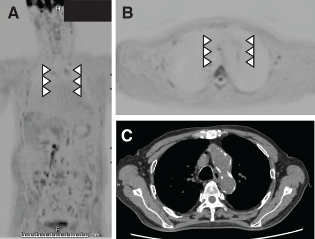
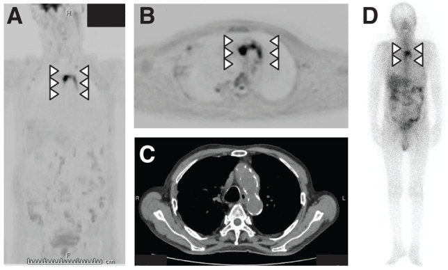
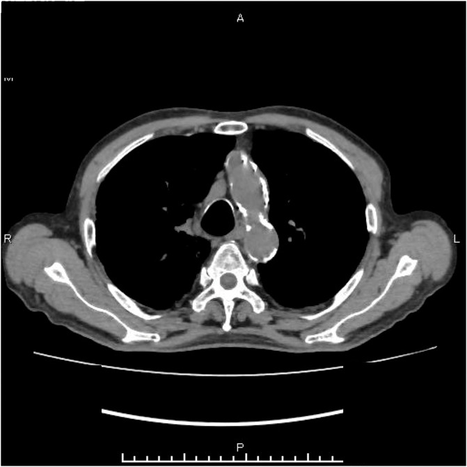
Diffusion-weighted whole-body imaging with background body signal suppression has been used to diagnose fever of unknown origin. An 86-year-old man who underwent bile duct jejunostomy for bile duct cancer presented with fever (body temperature, 40°C). Escherichia coli was detected in blood cultures. Diffusion-weighted whole-body imaging with background body signal suppression revealed accumulation in the aortic arch. Therefore, infectious aortitis secondary to retrograde cholangitis was diagnosed. The patient was treated with antibiotics, and the aortic arch accumulation disappeared. Diffusion-weighted whole-body imaging with background body signal suppression is a useful modality for diagnosing vasculitis and assessing treatment effectiveness.

 求助内容:
求助内容: 应助结果提醒方式:


