David Gergely Kovacs, Marianne Aznar, Marcel Van Herk, Iskandar Mohamed, James Price, Claes Nøhr Ladefoged, Barbara Malene Fischer, Flemming Littrup Andersen, Andrew McPartlin, Eliana M Vasquez Osorio, Azadeh Abravan
{"title":"深度学习衍生的18F-FDG PET/CT δ生物标志物在头颈癌局部区域控制中的外部验证。","authors":"David Gergely Kovacs, Marianne Aznar, Marcel Van Herk, Iskandar Mohamed, James Price, Claes Nøhr Ladefoged, Barbara Malene Fischer, Flemming Littrup Andersen, Andrew McPartlin, Eliana M Vasquez Osorio, Azadeh Abravan","doi":"10.2340/1651-226X.2025.43977","DOIUrl":null,"url":null,"abstract":"<p><strong>Background and purpose: </strong>Delta biomarkers that reflect changes in tumour burden over time can support personalised follow-up in head and neck cancer. However, their clinical use can be limited by the need for manual image segmentation. This study externally evaluates a deep learning model for automatic determination of volume change from serial 18F-fluorodeoxyglucose (18F-FDG) positron emission tomography/computed tomography (PET/CT) scans to stratify patients by loco-regional outcome. Patient/material and methods: An externally developed deep learning algorithm for tumour segmentation was applied to pre- and post-radiotherapy (RT, with or without concomitant chemoradiotherapy) PET/CT scans of 50 consecutive head and neck cancer patients from The Christie NHS Foundation Trust, UK. The model, originally trained on pre-treatment scans from a different institution, was deployed to derive tumour volumes at both time points. The AI-derived change in tumour volume (ΔPET-Gross tumour volume (GTV)) was calculated for each patient. Kaplan-Meier analysis assessed loco-regional control based on ΔPET-GTV, dichotomised at the cohort median. In a separate secondary analysis confined to the pre‑treatment scans, a radiation oncologist qualitatively evaluated the AI‑generated PET‑GTV contours.</p><p><strong>Results: </strong>Patients with higher ΔPET-GTV (i.e. greater tumour shrinkage) had significantly improved loco-regional control (log-rank p = 0.02). At 2 years, control was 94.1% (95% CI: 83.6-100%) vs. 53.6% (95% CI: 32.2-89.1%). Only one of nine failures occurred in the high ΔPET-GTV group. Clinician review found AI volumes acceptable for planning in 78% of cases. In two cases, the algorithm identified oropharyngeal primaries on pre-treatment PET-CT before clinical identification.</p><p><strong>Interpretation: </strong>Deep learning-derived ΔPET-GTV may support clinically meaningful assessment of post-treatment disease status and risk stratification, offering a scalable alternative to manual segmentation in PET/CT follow-up.</p>","PeriodicalId":7110,"journal":{"name":"Acta Oncologica","volume":"64 ","pages":"1143-1151"},"PeriodicalIF":2.7000,"publicationDate":"2025-08-30","publicationTypes":"Journal Article","fieldsOfStudy":null,"isOpenAccess":false,"openAccessPdf":"https://www.ncbi.nlm.nih.gov/pmc/articles/PMC12406743/pdf/","citationCount":"0","resultStr":"{\"title\":\"External validation of deep learning-derived 18F-FDG PET/CT delta biomarkers for loco-regional control in head and neck cancer.\",\"authors\":\"David Gergely Kovacs, Marianne Aznar, Marcel Van Herk, Iskandar Mohamed, James Price, Claes Nøhr Ladefoged, Barbara Malene Fischer, Flemming Littrup Andersen, Andrew McPartlin, Eliana M Vasquez Osorio, Azadeh Abravan\",\"doi\":\"10.2340/1651-226X.2025.43977\",\"DOIUrl\":null,\"url\":null,\"abstract\":\"<p><strong>Background and purpose: </strong>Delta biomarkers that reflect changes in tumour burden over time can support personalised follow-up in head and neck cancer. However, their clinical use can be limited by the need for manual image segmentation. This study externally evaluates a deep learning model for automatic determination of volume change from serial 18F-fluorodeoxyglucose (18F-FDG) positron emission tomography/computed tomography (PET/CT) scans to stratify patients by loco-regional outcome. Patient/material and methods: An externally developed deep learning algorithm for tumour segmentation was applied to pre- and post-radiotherapy (RT, with or without concomitant chemoradiotherapy) PET/CT scans of 50 consecutive head and neck cancer patients from The Christie NHS Foundation Trust, UK. The model, originally trained on pre-treatment scans from a different institution, was deployed to derive tumour volumes at both time points. The AI-derived change in tumour volume (ΔPET-Gross tumour volume (GTV)) was calculated for each patient. Kaplan-Meier analysis assessed loco-regional control based on ΔPET-GTV, dichotomised at the cohort median. In a separate secondary analysis confined to the pre‑treatment scans, a radiation oncologist qualitatively evaluated the AI‑generated PET‑GTV contours.</p><p><strong>Results: </strong>Patients with higher ΔPET-GTV (i.e. greater tumour shrinkage) had significantly improved loco-regional control (log-rank p = 0.02). At 2 years, control was 94.1% (95% CI: 83.6-100%) vs. 53.6% (95% CI: 32.2-89.1%). Only one of nine failures occurred in the high ΔPET-GTV group. Clinician review found AI volumes acceptable for planning in 78% of cases. In two cases, the algorithm identified oropharyngeal primaries on pre-treatment PET-CT before clinical identification.</p><p><strong>Interpretation: </strong>Deep learning-derived ΔPET-GTV may support clinically meaningful assessment of post-treatment disease status and risk stratification, offering a scalable alternative to manual segmentation in PET/CT follow-up.</p>\",\"PeriodicalId\":7110,\"journal\":{\"name\":\"Acta Oncologica\",\"volume\":\"64 \",\"pages\":\"1143-1151\"},\"PeriodicalIF\":2.7000,\"publicationDate\":\"2025-08-30\",\"publicationTypes\":\"Journal Article\",\"fieldsOfStudy\":null,\"isOpenAccess\":false,\"openAccessPdf\":\"https://www.ncbi.nlm.nih.gov/pmc/articles/PMC12406743/pdf/\",\"citationCount\":\"0\",\"resultStr\":null,\"platform\":\"Semanticscholar\",\"paperid\":null,\"PeriodicalName\":\"Acta Oncologica\",\"FirstCategoryId\":\"3\",\"ListUrlMain\":\"https://doi.org/10.2340/1651-226X.2025.43977\",\"RegionNum\":3,\"RegionCategory\":\"医学\",\"ArticlePicture\":[],\"TitleCN\":null,\"AbstractTextCN\":null,\"PMCID\":null,\"EPubDate\":\"\",\"PubModel\":\"\",\"JCR\":\"Q3\",\"JCRName\":\"ONCOLOGY\",\"Score\":null,\"Total\":0}","platform":"Semanticscholar","paperid":null,"PeriodicalName":"Acta Oncologica","FirstCategoryId":"3","ListUrlMain":"https://doi.org/10.2340/1651-226X.2025.43977","RegionNum":3,"RegionCategory":"医学","ArticlePicture":[],"TitleCN":null,"AbstractTextCN":null,"PMCID":null,"EPubDate":"","PubModel":"","JCR":"Q3","JCRName":"ONCOLOGY","Score":null,"Total":0}
引用次数: 0
摘要
背景和目的:反映肿瘤负荷随时间变化的Delta生物标志物可以支持头颈癌的个性化随访。然而,它们的临床应用可能受到人工图像分割的限制。本研究外部评估了一种深度学习模型,用于自动确定18f -氟脱氧葡萄糖(18F-FDG)正电子发射断层扫描/计算机断层扫描(PET/CT)扫描的体积变化,从而根据局部区域结果对患者进行分层。患者/材料和方法:外部开发的肿瘤分割深度学习算法应用于放疗前后(RT,伴或不伴放化疗)PET/CT扫描50例来自英国克里斯蒂NHS基金会信托的连续头颈癌患者。该模型最初是根据来自不同机构的治疗前扫描进行训练的,用于在两个时间点计算肿瘤体积。计算每位患者人工智能引起的肿瘤体积变化(ΔPET-Gross肿瘤体积(GTV))。Kaplan-Meier分析评估了基于ΔPET-GTV的局部区域控制,在队列中位数处进行二分。在一项单独的二次分析中,一名放射肿瘤学家定性地评估了人工智能生成的PET - GTV轮廓。结果:ΔPET-GTV较高的患者(即肿瘤缩小较大)的局部-区域控制显著改善(log-rank p = 0.02)。2年时,对照为94.1% (95% CI: 83.6-100%) vs. 53.6% (95% CI: 32.2-89.1%)。9次失败中只有1次发生在高ΔPET-GTV组。临床医生审查发现,在78%的病例中,人工智能体积可用于计划。在两个病例中,该算法在临床鉴定前先通过治疗前的PET-CT识别口咽原发病灶。解释:深度学习衍生的ΔPET-GTV可能支持治疗后疾病状态和风险分层的临床有意义的评估,为PET/CT随访中的人工分割提供可扩展的替代方案。
External validation of deep learning-derived 18F-FDG PET/CT delta biomarkers for loco-regional control in head and neck cancer.
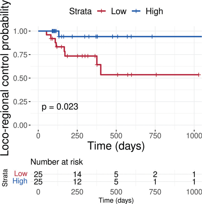
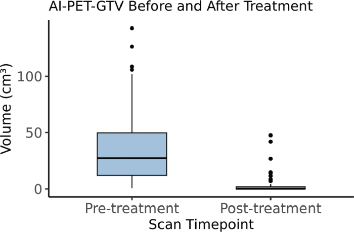
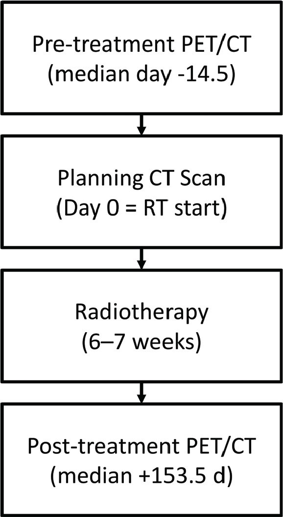
Background and purpose: Delta biomarkers that reflect changes in tumour burden over time can support personalised follow-up in head and neck cancer. However, their clinical use can be limited by the need for manual image segmentation. This study externally evaluates a deep learning model for automatic determination of volume change from serial 18F-fluorodeoxyglucose (18F-FDG) positron emission tomography/computed tomography (PET/CT) scans to stratify patients by loco-regional outcome. Patient/material and methods: An externally developed deep learning algorithm for tumour segmentation was applied to pre- and post-radiotherapy (RT, with or without concomitant chemoradiotherapy) PET/CT scans of 50 consecutive head and neck cancer patients from The Christie NHS Foundation Trust, UK. The model, originally trained on pre-treatment scans from a different institution, was deployed to derive tumour volumes at both time points. The AI-derived change in tumour volume (ΔPET-Gross tumour volume (GTV)) was calculated for each patient. Kaplan-Meier analysis assessed loco-regional control based on ΔPET-GTV, dichotomised at the cohort median. In a separate secondary analysis confined to the pre‑treatment scans, a radiation oncologist qualitatively evaluated the AI‑generated PET‑GTV contours.
Results: Patients with higher ΔPET-GTV (i.e. greater tumour shrinkage) had significantly improved loco-regional control (log-rank p = 0.02). At 2 years, control was 94.1% (95% CI: 83.6-100%) vs. 53.6% (95% CI: 32.2-89.1%). Only one of nine failures occurred in the high ΔPET-GTV group. Clinician review found AI volumes acceptable for planning in 78% of cases. In two cases, the algorithm identified oropharyngeal primaries on pre-treatment PET-CT before clinical identification.
Interpretation: Deep learning-derived ΔPET-GTV may support clinically meaningful assessment of post-treatment disease status and risk stratification, offering a scalable alternative to manual segmentation in PET/CT follow-up.
期刊介绍:
Acta Oncologica is a journal for the clinical oncologist and accepts articles within all fields of clinical cancer research. Articles on tumour pathology, experimental oncology, radiobiology, cancer epidemiology and medical radio physics are also welcome, especially if they have a clinical aim or interest. Scientific articles on cancer nursing and psychological or social aspects of cancer are also welcomed. Extensive material may be published as Supplements, for which special conditions apply.
 求助内容:
求助内容: 应助结果提醒方式:


