Xiaomei Zhong , Huan Lin , Rong Zhang , Yunlang She , Xiaobo Chen , Wei Zhao , Shiwei Luo , Entao Liu , Qingyu Liu , Jun Liu , Xin Chen , Zaiyi Liu
{"title":"IB-IIA期非小细胞肺癌多特征风险分层预测模型的建立:一项多中心分析","authors":"Xiaomei Zhong , Huan Lin , Rong Zhang , Yunlang She , Xiaobo Chen , Wei Zhao , Shiwei Luo , Entao Liu , Qingyu Liu , Jun Liu , Xin Chen , Zaiyi Liu","doi":"10.1016/j.ejrad.2025.112379","DOIUrl":null,"url":null,"abstract":"<div><h3>Objective</h3><div>This study aimed to develop a comprehensive risk stratification model for stage IB-IIA non-small cell lung cancer (NSCLC) by integrating clinicopathological data with pre-treatment CT imaging.</div></div><div><h3>Methods</h3><div>This retrospective study included three independent cohorts of patients with stage IB-IIA NSCLC for model development and validation (Training: n = 370; Internal validation: n = 120; External validation: n = 70). Disease-free survival (DFS) was the primary endpoint. Radiomics features were extracted from both tumoral and peritumoral regions of CT images to construct a radiomics model. A ResNet50-based deep learning architecture was adopted to develop a deep learning model using CT imaging data. Logistic regression was used to identify significant clinicopathological factors. These components were integrated into a multi-feature combined model (CRD model) that utilized clinicopathological, radiomics, and deep learning features for DFS prediction. Model interpretability was assessed using the SHapley Additive exPlanations (SHAP) method.</div></div><div><h3>Results</h3><div>The combined CRD model demonstrated superior performance in predicting DFS, achieving areas under the curve (AUC) of 0.865, 0.798, and 0.803 in the training, internal validation, and external validation cohorts, respectively. Patients were stratified into high- and low-risk groups using the CRD model, and in the external validation cohort, the hazard ratio (HR) for high-risk patients was 17.509, with a C-index of 0.73. SHAP analysis revealed that radiomics features contributed most significantly to the performance of the CRD model.</div></div><div><h3>Conclusions</h3><div>The multi-feature combined model effectively predicts DFS and identifies high-risk patients with stage IB-IIA NSCLC. It could facilitate personalized postoperative treatment strategies, improving patient outcomes.</div></div>","PeriodicalId":12063,"journal":{"name":"European Journal of Radiology","volume":"192 ","pages":"Article 112379"},"PeriodicalIF":3.3000,"publicationDate":"2025-08-24","publicationTypes":"Journal Article","fieldsOfStudy":null,"isOpenAccess":false,"openAccessPdf":"","citationCount":"0","resultStr":"{\"title\":\"Development of a multi-feature predictive model for risk stratification in stage IB-IIA non-small cell lung cancer: a multicenter analysis\",\"authors\":\"Xiaomei Zhong , Huan Lin , Rong Zhang , Yunlang She , Xiaobo Chen , Wei Zhao , Shiwei Luo , Entao Liu , Qingyu Liu , Jun Liu , Xin Chen , Zaiyi Liu\",\"doi\":\"10.1016/j.ejrad.2025.112379\",\"DOIUrl\":null,\"url\":null,\"abstract\":\"<div><h3>Objective</h3><div>This study aimed to develop a comprehensive risk stratification model for stage IB-IIA non-small cell lung cancer (NSCLC) by integrating clinicopathological data with pre-treatment CT imaging.</div></div><div><h3>Methods</h3><div>This retrospective study included three independent cohorts of patients with stage IB-IIA NSCLC for model development and validation (Training: n = 370; Internal validation: n = 120; External validation: n = 70). Disease-free survival (DFS) was the primary endpoint. Radiomics features were extracted from both tumoral and peritumoral regions of CT images to construct a radiomics model. A ResNet50-based deep learning architecture was adopted to develop a deep learning model using CT imaging data. Logistic regression was used to identify significant clinicopathological factors. These components were integrated into a multi-feature combined model (CRD model) that utilized clinicopathological, radiomics, and deep learning features for DFS prediction. Model interpretability was assessed using the SHapley Additive exPlanations (SHAP) method.</div></div><div><h3>Results</h3><div>The combined CRD model demonstrated superior performance in predicting DFS, achieving areas under the curve (AUC) of 0.865, 0.798, and 0.803 in the training, internal validation, and external validation cohorts, respectively. Patients were stratified into high- and low-risk groups using the CRD model, and in the external validation cohort, the hazard ratio (HR) for high-risk patients was 17.509, with a C-index of 0.73. SHAP analysis revealed that radiomics features contributed most significantly to the performance of the CRD model.</div></div><div><h3>Conclusions</h3><div>The multi-feature combined model effectively predicts DFS and identifies high-risk patients with stage IB-IIA NSCLC. It could facilitate personalized postoperative treatment strategies, improving patient outcomes.</div></div>\",\"PeriodicalId\":12063,\"journal\":{\"name\":\"European Journal of Radiology\",\"volume\":\"192 \",\"pages\":\"Article 112379\"},\"PeriodicalIF\":3.3000,\"publicationDate\":\"2025-08-24\",\"publicationTypes\":\"Journal Article\",\"fieldsOfStudy\":null,\"isOpenAccess\":false,\"openAccessPdf\":\"\",\"citationCount\":\"0\",\"resultStr\":null,\"platform\":\"Semanticscholar\",\"paperid\":null,\"PeriodicalName\":\"European Journal of Radiology\",\"FirstCategoryId\":\"3\",\"ListUrlMain\":\"https://www.sciencedirect.com/science/article/pii/S0720048X25004656\",\"RegionNum\":3,\"RegionCategory\":\"医学\",\"ArticlePicture\":[],\"TitleCN\":null,\"AbstractTextCN\":null,\"PMCID\":null,\"EPubDate\":\"\",\"PubModel\":\"\",\"JCR\":\"Q1\",\"JCRName\":\"RADIOLOGY, NUCLEAR MEDICINE & MEDICAL IMAGING\",\"Score\":null,\"Total\":0}","platform":"Semanticscholar","paperid":null,"PeriodicalName":"European Journal of Radiology","FirstCategoryId":"3","ListUrlMain":"https://www.sciencedirect.com/science/article/pii/S0720048X25004656","RegionNum":3,"RegionCategory":"医学","ArticlePicture":[],"TitleCN":null,"AbstractTextCN":null,"PMCID":null,"EPubDate":"","PubModel":"","JCR":"Q1","JCRName":"RADIOLOGY, NUCLEAR MEDICINE & MEDICAL IMAGING","Score":null,"Total":0}
Development of a multi-feature predictive model for risk stratification in stage IB-IIA non-small cell lung cancer: a multicenter analysis
Objective
This study aimed to develop a comprehensive risk stratification model for stage IB-IIA non-small cell lung cancer (NSCLC) by integrating clinicopathological data with pre-treatment CT imaging.
Methods
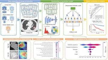
This retrospective study included three independent cohorts of patients with stage IB-IIA NSCLC for model development and validation (Training: n = 370; Internal validation: n = 120; External validation: n = 70). Disease-free survival (DFS) was the primary endpoint. Radiomics features were extracted from both tumoral and peritumoral regions of CT images to construct a radiomics model. A ResNet50-based deep learning architecture was adopted to develop a deep learning model using CT imaging data. Logistic regression was used to identify significant clinicopathological factors. These components were integrated into a multi-feature combined model (CRD model) that utilized clinicopathological, radiomics, and deep learning features for DFS prediction. Model interpretability was assessed using the SHapley Additive exPlanations (SHAP) method.
Results
The combined CRD model demonstrated superior performance in predicting DFS, achieving areas under the curve (AUC) of 0.865, 0.798, and 0.803 in the training, internal validation, and external validation cohorts, respectively. Patients were stratified into high- and low-risk groups using the CRD model, and in the external validation cohort, the hazard ratio (HR) for high-risk patients was 17.509, with a C-index of 0.73. SHAP analysis revealed that radiomics features contributed most significantly to the performance of the CRD model.
Conclusions
The multi-feature combined model effectively predicts DFS and identifies high-risk patients with stage IB-IIA NSCLC. It could facilitate personalized postoperative treatment strategies, improving patient outcomes.
期刊介绍:
European Journal of Radiology is an international journal which aims to communicate to its readers, state-of-the-art information on imaging developments in the form of high quality original research articles and timely reviews on current developments in the field.
Its audience includes clinicians at all levels of training including radiology trainees, newly qualified imaging specialists and the experienced radiologist. Its aim is to inform efficient, appropriate and evidence-based imaging practice to the benefit of patients worldwide.