使用简短的脑MRI扫描协议检测关键发现的准确性是人工智能驱动的实时扫描协议适应的先决条件
IF 3.3
3区 医学
Q1 RADIOLOGY, NUCLEAR MEDICINE & MEDICAL IMAGING
引用次数: 0
摘要
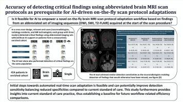
目的评估人工智能工具和相关放射学专业人员在使用简短的脑MRI扫描协议检测脑梗死、颅内出血和肿瘤方面的表现,作为基于实时成像结果动态选择额外成像序列的人工智能驱动工作流程的先决条件。材料与方法对来自丹麦四家医院的成人常规脑MRI扫描进行回顾性、连续充实队列研究。三名神经放射科顾问医师、三名放射科住院医师、三名磁共振技术专家和一种人工智能工具使用简化的3序列方案(DWI、SWI/T2*-GRE、T2- flair)或4序列方案(DWI、SWI/T2*-GRE、T2- flair、T1W)在非重叠的三向分裂交叉设计中检测脑梗死、出血和肿瘤。参考结果是根据放射学报告和独立图像审查建立的。结果共纳入414例患者(57岁±19238例男性),其中确诊脑梗死65例,出血65例,肿瘤65例。人工智能工具对脑梗死、出血和肿瘤的检测灵敏度分别为94%(65次扫描中的61次,95% CI:85 - 98%)、82%(65次扫描中的53次,95% CI:70 - 90%)和74%(65次扫描中的48次,95% CI:61 - 84%),相应的特异性为86%(349次扫描中的300次,95% CI:82 - 89%)、84%(349次扫描中的293次,95% CI:80 - 88%)和62%(349次扫描中的217次,95% CI:57 - 67%)。该工具的灵敏度可与神经放射学家使用简化扫描协议的灵敏度相媲美,并且在肿瘤检测方面超过了MR技术专家,尽管其特异性较低。结论自动化实时扫描适应的初步步骤是可行的,并且可以潜在地提高检测灵敏度,平衡与当前护理标准相比降低的特异性。本文章由计算机程序翻译,如有差异,请以英文原文为准。
Accuracy of detecting critical findings using abbreviated brain MRI scan protocols as a prerequisite for AI-driven on-the-fly scan protocol adaptation
Purpose
To evaluate the performance of an AI tool and relevant radiology professionals in detecting brain infarcts, intracranial hemorrhages, and tumors using abbreviated brain MRI scan protocols as prerequisite for an AI-driven workflow that dynamically selects additional imaging sequences based on real-time imaging findings.
Materials and methods
A retrospective, consecutively enriched cohort of routine adult brain MRI scans from four Danish hospitals was constructed. Three consultant neuroradiologists, three radiology residents, three MR technologists, and an AI tool detected brain infarcts, hemorrhages, and tumors using an abbreviated 3-sequence protocol (DWI, SWI/T2*-GRE, T2-FLAIR) or 4-sequence protocol (DWI, SWI/T2*-GRE, T2-FLAIR, T1W) in a non-overlapping three-way split cross-over design. Reference findings were established from radiological reports and independent image reviews.
Results
A total of 414 patients were included (57 years ± 19, 238 men) with 65 confirmed brain infarcts, 65 hemorrhages, and 65 tumors. The AI tool achieved detection sensitivities of 94 % (61 of 65 scans, 95 %CI:85–98 %), 82 % (53 of 65 scans, 95 %CI:70–90 %), and 74 % (48 of 65 scans, 95 %CI:61–84 %) for brain infarcts, hemorrhages, and tumors, respectively, and corresponding specificities of 86 % (300 of 349 scans, 95 %CI:82–89 %), 84 % (293 of 349 scans, 95 %CI:80–88 %), and 62 % (217 of 349 scans, 95 %CI:57–67 %). The tool achieved sensitivities comparable to those of neuroradiologists using abbreviated scan protocols, and it surpassed MR technologists in tumor detection, though its specificities were lower.
Conclusion
The initial steps towards automated real-time scan adaptation are feasible and can potentially improve detection sensitivity, balancing reduced specificities compared to the current standard of care.
求助全文
通过发布文献求助,成功后即可免费获取论文全文。
去求助
来源期刊

CiteScore
6.70
自引率
3.00%
发文量
398
审稿时长
42 days
期刊介绍:
European Journal of Radiology is an international journal which aims to communicate to its readers, state-of-the-art information on imaging developments in the form of high quality original research articles and timely reviews on current developments in the field.
Its audience includes clinicians at all levels of training including radiology trainees, newly qualified imaging specialists and the experienced radiologist. Its aim is to inform efficient, appropriate and evidence-based imaging practice to the benefit of patients worldwide.
 求助内容:
求助内容: 应助结果提醒方式:


