股骨冠状面弓形:1313根股骨的计算机断层分析
IF 3.6
2区 医学
Q2 ENDOCRINOLOGY & METABOLISM
引用次数: 0
摘要
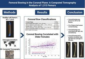
对矢状面股弓形的认识导致了植入物设计和放置技术的优化。然而,对冠状面股弓形的了解甚少。本研究的目的是描述股骨冠状弯曲以及人口统计学特征如何与弯曲相关。方法对Stryker骨科建模与分析数据库中1313根左股骨的三维模型进行分析。冠状弓以厘米(cm)为曲率半径测量,分为直(>300 cm)、轻度至中度(>300 cm)和重度(<150 cm)。进行单变量分析以检测人口统计学和冠状动脉弯曲之间的关联。还进行了多变量分析,包括性别特异性Spearman相关系数。结果236例(18%)股骨发生轻至中度外侧弯曲,46例(4%)股骨发生严重外侧弯曲,35例(3%)股骨发生轻至中度内侧弯曲,2例(1%)股骨发生严重内侧弯曲。单变量分析显示冠状动脉弯曲与年龄(p < 0.001)、性别(p < 0.001)和种族(p = 0.002)有显著相关性,但与体重指数无关(p = 0.54)。多变量分析发现,年龄与冠状动脉弯曲的相关性在女性中呈中等正相关(Spearman’s Rho = 0.42, p < 0.001),而在男性中呈弱正相关(Spearman’s Rho = 0.21, p < 0.001)。结论1313根股骨的三维分析表明,24%的股骨有冠状弓形,这与年龄、性别和种族有显著相关性。冠状弓形与年龄呈正相关,且在女性中更为明显。通过承认股骨形态的变化,这些发现强调了在术前计划期间检查受伤和未受伤股骨的多平面x线片的实用性。本文章由计算机程序翻译,如有差异,请以英文原文为准。
Femoral bowing in the coronal plane: A computed tomography analysis of 1313 femurs
Introduction
Appreciation of femoral bowing in the sagittal plane has led to optimization of implant design and placement techniques. However, little is known about femoral bowing in the coronal plane. The objective of this study was to describe coronal bowing of the femur and how demographic characteristics were associated with bowing.
Methods
Three-dimensional models of 1313 left femurs taken from the Stryker Orthopaedic Modeling and Analytics database were analyzed. Coronal bowing was measured via radius of curvature in centimeters (cm) and classified as straight (>300 cm), mild-to-moderate (150 to 300 cm), and severe (<150 cm). Univariable analyses were performed to detect associations between demographics and coronal bowing. Multivariable analyses including sex-specific Spearman correlation coefficients were also undertaken.
Results
In total, 236 femurs (18 %) had mild-to-moderate lateral bowing, 46 (4 %) had severe lateral bowing, 35 (3 %) had mild-to-moderate medial bowing, and 2 (<1 %) had severe medial bowing. Univariable analyses revealed coronal bowing had significant associations with age (p < 0.001), sex (p < 0.001), and ethnicity (p = 0.002) but not with body mass index (p = 0.54). Multivariable analyses found the correlation of age with coronal bowing to be moderate positive in females (Spearman's Rho = 0.42, p < 0.001) compared to weak positive in males (Spearman's Rho = 0.21, p < 0.001).
Conclusions
Three-dimensional analysis of 1313 femurs demonstrated that 24 % of femurs had coronal bowing which had significant associations with age, sex, and ethnicity. Coronal bowing had a positive correlation with age that was stronger in females. By acknowledging variations in femoral morphology, these findings highlight the utility of examining radiographs of the injured and uninjured femurs in multiple planes during preoperative planning.
求助全文
通过发布文献求助,成功后即可免费获取论文全文。
去求助
来源期刊

Bone
医学-内分泌学与代谢
CiteScore
8.90
自引率
4.90%
发文量
264
审稿时长
30 days
期刊介绍:
BONE is an interdisciplinary forum for the rapid publication of original articles and reviews on basic, translational, and clinical aspects of bone and mineral metabolism. The Journal also encourages submissions related to interactions of bone with other organ systems, including cartilage, endocrine, muscle, fat, neural, vascular, gastrointestinal, hematopoietic, and immune systems. Particular attention is placed on the application of experimental studies to clinical practice.
 求助内容:
求助内容: 应助结果提醒方式:


