FastDIP:一种加速无监督低计数PET图像重建的有效方法
IF 4.9
2区 医学
Q1 ENGINEERING, BIOMEDICAL
Computerized Medical Imaging and Graphics
Pub Date : 2025-08-21
DOI:10.1016/j.compmedimag.2025.102639
引用次数: 0
摘要
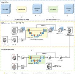
无监督深度学习方法可以在不需要大规模数据集的情况下提高正电子发射断层扫描(PET)图像的图像质量。然而,这些方法通常需要为每个患者训练一个不同的网络,使得重建过程非常耗时,并且限制了它们的临床适用性。在本文中,我们的研究目标是为无监督PET图像重建开发一种高效的无监督学习框架,以满足临床对实时成像能力的需求。方法:在本研究中,我们提出了FastDIP,一种用于无监督低计数PET图像重建的高效学习方法。FastDIP采用两阶段重建过程,首先是快速的粗重建,然后是详细的精细重建。采用像素shuffle下采样方法对PET图像进行压缩,实现快速粗重建。此外,一个小波去噪的PET图像作为输入,取代传统的解剖图像。我们还结合了预训练技术来加速网络收敛。结果:FastDIP在模拟18F-AV45脑数据集、临床18F-FDG脑数据集和临床68Ga-PSMA体数据集上的疗效进行了评估。FastDIP与深度图像先验(DIP)、条件深度图像先验(CDIP)、引导深度图像先验(GDIP)、自监督预训练DIP (SPDIP)、群体预训练DIP (PPDIP)和各种消融方法进行了比较。对于18F-AV45数据集,FastDIP在不同计数水平上仅使用11%的训练时间就获得了比DIP更好的图像质量。在18F-FDG数据集中,该方法在2.2 min内实现了最低的归一化均方误差和最高的结构相似性,优于DIP (10.7 min)、CDIP (7.5 min)、GDIP (9.8 min)、SPDIP (166.7 min)和PPDIP (166.7 min)。对于68Ga-PSMA数据集,FastDIP在2.4 min内实现了最高的噪比和SUVmax,超过了DIP (10.7 min)、CDIP (32.0 min)、GDIP (32.7 min)、SPDIP (16.7 min)和PPDIP (37.5 min)。结论:FastDIP是一种有效的无监督低计数PET图像重建方法,可显著减少网络训练时间,显著提高图像恢复性能。本文章由计算机程序翻译,如有差异,请以英文原文为准。
FastDIP: An effective approach for accelerating unsupervised low-count PET image reconstruction
Introduction:
Unsupervised deep learning methods can improve the image quality of positron emission tomography (PET) images without the need for large-scale datasets. However, these approaches typically require training a distinct network for each patient, making the reconstruction process extremely time-consuming and limiting their clinical applicability. In this paper, our research objective is to develop an efficient unsupervised learning framework for unsupervised PET image reconstruction, in order to fulfill the clinical requirement for real-time imaging capabilities.
Methods:
In this study, we present FastDIP, an efficient learning method for unsupervised low-count PET image reconstruction. FastDIP employs a two-stage reconstruction process, beginning with a rapid coarse reconstruction followed by a detailed fine reconstruction. The pixel-shuffle downsampling method is utilized to compress PET images and facilitate quick coarse reconstruction. Additionally, a wavelet-denoised PET image serves as input, replacing the traditional anatomical images. We also incorporate pre-training techniques to accelerate network convergence.
Results:
The efficacy of FastDIP was evaluated on simulated 18F-AV45 brain datasets, as well as clinical 18F-FDG brain and clinical 68Ga-PSMA body datasets. FastDIP was compared to Deep Image Prior (DIP), Conditional Deep Image Prior (CDIP), Guided Deep Image Prior (GDIP), Self-supervised Pre-training DIP (SPDIP), Population Pre-training DIP (PPDIP) and various ablation methods. For the 18F-AV45 dataset, FastDIP achieved better image quality than DIP using only 11% training time of them across different count levels. In the 18F-FDG dataset, it achieved the lowest normalized mean square error and the highest structural similarity in just 2.2 min, outperforming DIP (10.7 min), CDIP (7.5 min), GDIP (9.8 min), SPDIP (166.7 min) and PPDIP (166.7 min). For the 68Ga-PSMA dataset, FastDIP achieved the highest contrast-to-noise ratio and SUVmax in 2.4 min, surpassing DIP (10.7 min), CDIP (32.0 min) , GDIP (32.7 min), SPDIP (16.7 min) and PPDIP (37.5 min).
Conclusion:
FastDIP is an efficient approach for unsupervised low-count PET image reconstruction that significantly reduces the network training time and markedly enhances image restoration performance.
求助全文
通过发布文献求助,成功后即可免费获取论文全文。
去求助
来源期刊

CiteScore
10.70
自引率
3.50%
发文量
71
审稿时长
26 days
期刊介绍:
The purpose of the journal Computerized Medical Imaging and Graphics is to act as a source for the exchange of research results concerning algorithmic advances, development, and application of digital imaging in disease detection, diagnosis, intervention, prevention, precision medicine, and population health. Included in the journal will be articles on novel computerized imaging or visualization techniques, including artificial intelligence and machine learning, augmented reality for surgical planning and guidance, big biomedical data visualization, computer-aided diagnosis, computerized-robotic surgery, image-guided therapy, imaging scanning and reconstruction, mobile and tele-imaging, radiomics, and imaging integration and modeling with other information relevant to digital health. The types of biomedical imaging include: magnetic resonance, computed tomography, ultrasound, nuclear medicine, X-ray, microwave, optical and multi-photon microscopy, video and sensory imaging, and the convergence of biomedical images with other non-imaging datasets.
 求助内容:
求助内容: 应助结果提醒方式:


