{"title":"EOS三维成像系统测量复发性髌骨脱位患者TT-TG距离的可靠性和可重复性。","authors":"Xian Zhang, Boming Zhao, Jiang Zheng, Bo Ren","doi":"10.1177/23259671251361493","DOIUrl":null,"url":null,"abstract":"<p><strong>Background: </strong>In clinical practice, preoperative radiographic assessment of the tibial tuberosity-trochlear groove (TT-TG) distance in patients with recurrent patellar dislocation (RPD) typically relies on conventional computed tomography (CT). A novel EOS imaging system may be a new option for measuring the TT-TG, which provides less radiation and costly time to perform.</p><p><strong>Purpose: </strong>To determine the reliability and reproducibility of the EOS 3-dimensional (3D) imaging system to measure TT-TG values.</p><p><strong>Study design: </strong>Cohort study (Diagnosis); Level of evidence, 2.</p><p><strong>Methods: </strong>Data from 34 patients (36 knees) diagnosed with RPD in our sports medicine department were retrospectively analyzed. Two senior radiologists independently measured the alignment of the patients' lower limbs via EOS images. The TT-TG values measured by EOS 3D images and CT scans were compared. To evaluate intrareader reliability, measurements were repeated on 2 separate occasions ≥3 days apart and intraclass correlation coefficients (ICCs) and Bland-Altman plots were calculated.</p><p><strong>Results: </strong>When lower-limb alignment was measured with EOS, there were no statistically significant differences in the femoral and tibial lengths (<i>P</i> = .87 and <i>P</i> = .78, respectively), femoral offset (<i>P</i> = .83), knee joint varus and valgus angles (<i>P</i> = .73), femoral and tibial mechanical angle (<i>P</i> = .70 and <i>P</i> = .83, respectively), and femorotibial torsion angles (<i>P</i> = .82) measured by the 2 radiologists. Excellent intraobserver ICC (>0.75) was observed. The TT-TG measurements using EOS 3D were 18.4 ± 4.2 mm (radiologist 1) and 18.4 ± 3.5 mm (radiologist 2), while the corresponding values from CT were 19.2 ± 4.0 mm and 18.5 ± 3.7 mm, respectively. The measurements between EOS and CT showed good intrarater consistency (ICC > 0.7). Bland-Altman analysis revealed mean differences of 0.743 mm and 0.081 mm between the 2 methods, with >91% of data points falling within ±1.96 SD, indicating strong agreement between the 2 measurement techniques.</p><p><strong>Conclusion: </strong>Our study showed that the EOS 3D imaging system provides reliable and reproducible TT-TG measurements comparable with CT. This technology has the potential to serve as an alternative method for assessing lower-limb alignment and TT-TG values in patients with RPD.</p>","PeriodicalId":19646,"journal":{"name":"Orthopaedic Journal of Sports Medicine","volume":"13 8","pages":"23259671251361493"},"PeriodicalIF":2.5000,"publicationDate":"2025-08-13","publicationTypes":"Journal Article","fieldsOfStudy":null,"isOpenAccess":false,"openAccessPdf":"https://www.ncbi.nlm.nih.gov/pmc/articles/PMC12351149/pdf/","citationCount":"0","resultStr":"{\"title\":\"Reliability and Reproducibility of the EOS 3D Imaging System in Measuring TT-TG Distance in Patients With Recurrent Patellar Dislocation.\",\"authors\":\"Xian Zhang, Boming Zhao, Jiang Zheng, Bo Ren\",\"doi\":\"10.1177/23259671251361493\",\"DOIUrl\":null,\"url\":null,\"abstract\":\"<p><strong>Background: </strong>In clinical practice, preoperative radiographic assessment of the tibial tuberosity-trochlear groove (TT-TG) distance in patients with recurrent patellar dislocation (RPD) typically relies on conventional computed tomography (CT). A novel EOS imaging system may be a new option for measuring the TT-TG, which provides less radiation and costly time to perform.</p><p><strong>Purpose: </strong>To determine the reliability and reproducibility of the EOS 3-dimensional (3D) imaging system to measure TT-TG values.</p><p><strong>Study design: </strong>Cohort study (Diagnosis); Level of evidence, 2.</p><p><strong>Methods: </strong>Data from 34 patients (36 knees) diagnosed with RPD in our sports medicine department were retrospectively analyzed. Two senior radiologists independently measured the alignment of the patients' lower limbs via EOS images. The TT-TG values measured by EOS 3D images and CT scans were compared. To evaluate intrareader reliability, measurements were repeated on 2 separate occasions ≥3 days apart and intraclass correlation coefficients (ICCs) and Bland-Altman plots were calculated.</p><p><strong>Results: </strong>When lower-limb alignment was measured with EOS, there were no statistically significant differences in the femoral and tibial lengths (<i>P</i> = .87 and <i>P</i> = .78, respectively), femoral offset (<i>P</i> = .83), knee joint varus and valgus angles (<i>P</i> = .73), femoral and tibial mechanical angle (<i>P</i> = .70 and <i>P</i> = .83, respectively), and femorotibial torsion angles (<i>P</i> = .82) measured by the 2 radiologists. Excellent intraobserver ICC (>0.75) was observed. The TT-TG measurements using EOS 3D were 18.4 ± 4.2 mm (radiologist 1) and 18.4 ± 3.5 mm (radiologist 2), while the corresponding values from CT were 19.2 ± 4.0 mm and 18.5 ± 3.7 mm, respectively. The measurements between EOS and CT showed good intrarater consistency (ICC > 0.7). Bland-Altman analysis revealed mean differences of 0.743 mm and 0.081 mm between the 2 methods, with >91% of data points falling within ±1.96 SD, indicating strong agreement between the 2 measurement techniques.</p><p><strong>Conclusion: </strong>Our study showed that the EOS 3D imaging system provides reliable and reproducible TT-TG measurements comparable with CT. This technology has the potential to serve as an alternative method for assessing lower-limb alignment and TT-TG values in patients with RPD.</p>\",\"PeriodicalId\":19646,\"journal\":{\"name\":\"Orthopaedic Journal of Sports Medicine\",\"volume\":\"13 8\",\"pages\":\"23259671251361493\"},\"PeriodicalIF\":2.5000,\"publicationDate\":\"2025-08-13\",\"publicationTypes\":\"Journal Article\",\"fieldsOfStudy\":null,\"isOpenAccess\":false,\"openAccessPdf\":\"https://www.ncbi.nlm.nih.gov/pmc/articles/PMC12351149/pdf/\",\"citationCount\":\"0\",\"resultStr\":null,\"platform\":\"Semanticscholar\",\"paperid\":null,\"PeriodicalName\":\"Orthopaedic Journal of Sports Medicine\",\"FirstCategoryId\":\"3\",\"ListUrlMain\":\"https://doi.org/10.1177/23259671251361493\",\"RegionNum\":3,\"RegionCategory\":\"医学\",\"ArticlePicture\":[],\"TitleCN\":null,\"AbstractTextCN\":null,\"PMCID\":null,\"EPubDate\":\"2025/8/1 0:00:00\",\"PubModel\":\"eCollection\",\"JCR\":\"Q2\",\"JCRName\":\"ORTHOPEDICS\",\"Score\":null,\"Total\":0}","platform":"Semanticscholar","paperid":null,"PeriodicalName":"Orthopaedic Journal of Sports Medicine","FirstCategoryId":"3","ListUrlMain":"https://doi.org/10.1177/23259671251361493","RegionNum":3,"RegionCategory":"医学","ArticlePicture":[],"TitleCN":null,"AbstractTextCN":null,"PMCID":null,"EPubDate":"2025/8/1 0:00:00","PubModel":"eCollection","JCR":"Q2","JCRName":"ORTHOPEDICS","Score":null,"Total":0}
Reliability and Reproducibility of the EOS 3D Imaging System in Measuring TT-TG Distance in Patients With Recurrent Patellar Dislocation.
Background: In clinical practice, preoperative radiographic assessment of the tibial tuberosity-trochlear groove (TT-TG) distance in patients with recurrent patellar dislocation (RPD) typically relies on conventional computed tomography (CT). A novel EOS imaging system may be a new option for measuring the TT-TG, which provides less radiation and costly time to perform.
Purpose: To determine the reliability and reproducibility of the EOS 3-dimensional (3D) imaging system to measure TT-TG values.
Study design: Cohort study (Diagnosis); Level of evidence, 2.
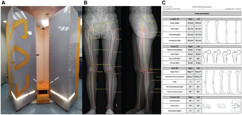
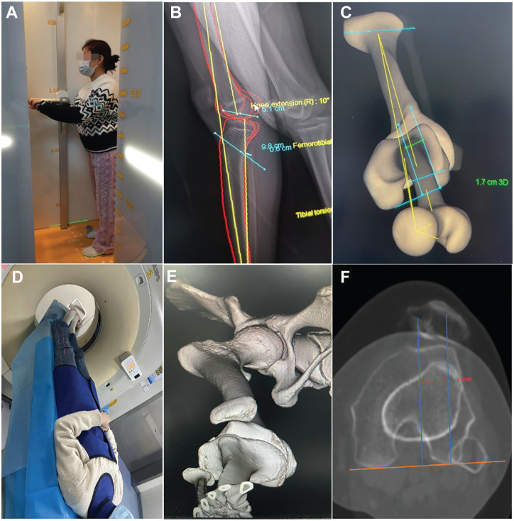
Methods: Data from 34 patients (36 knees) diagnosed with RPD in our sports medicine department were retrospectively analyzed. Two senior radiologists independently measured the alignment of the patients' lower limbs via EOS images. The TT-TG values measured by EOS 3D images and CT scans were compared. To evaluate intrareader reliability, measurements were repeated on 2 separate occasions ≥3 days apart and intraclass correlation coefficients (ICCs) and Bland-Altman plots were calculated.
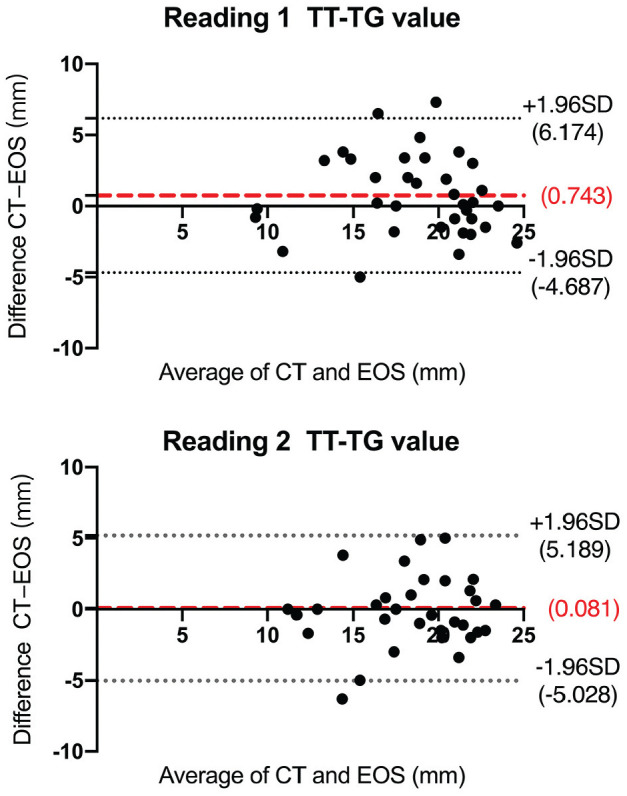
Results: When lower-limb alignment was measured with EOS, there were no statistically significant differences in the femoral and tibial lengths (P = .87 and P = .78, respectively), femoral offset (P = .83), knee joint varus and valgus angles (P = .73), femoral and tibial mechanical angle (P = .70 and P = .83, respectively), and femorotibial torsion angles (P = .82) measured by the 2 radiologists. Excellent intraobserver ICC (>0.75) was observed. The TT-TG measurements using EOS 3D were 18.4 ± 4.2 mm (radiologist 1) and 18.4 ± 3.5 mm (radiologist 2), while the corresponding values from CT were 19.2 ± 4.0 mm and 18.5 ± 3.7 mm, respectively. The measurements between EOS and CT showed good intrarater consistency (ICC > 0.7). Bland-Altman analysis revealed mean differences of 0.743 mm and 0.081 mm between the 2 methods, with >91% of data points falling within ±1.96 SD, indicating strong agreement between the 2 measurement techniques.
Conclusion: Our study showed that the EOS 3D imaging system provides reliable and reproducible TT-TG measurements comparable with CT. This technology has the potential to serve as an alternative method for assessing lower-limb alignment and TT-TG values in patients with RPD.
期刊介绍:
The Orthopaedic Journal of Sports Medicine (OJSM), developed by the American Orthopaedic Society for Sports Medicine (AOSSM), is a global, peer-reviewed, open access journal that combines the interests of researchers and clinical practitioners across orthopaedic sports medicine, arthroscopy, and knee arthroplasty.
Topics include original research in the areas of:
-Orthopaedic Sports Medicine, including surgical and nonsurgical treatment of orthopaedic sports injuries
-Arthroscopic Surgery (Shoulder/Elbow/Wrist/Hip/Knee/Ankle/Foot)
-Relevant translational research
-Sports traumatology/epidemiology
-Knee and shoulder arthroplasty
The OJSM also publishes relevant systematic reviews and meta-analyses.
This journal is a member of the Committee on Publication Ethics (COPE).

 求助内容:
求助内容: 应助结果提醒方式:


