基于计算机断层扫描/磁共振成像多模态图像融合的三维重建在腮腺肿瘤诊断与治疗中的应用。

IF 2.1 2区 医学 Q2 DENTISTRY, ORAL SURGERY & MEDICINE
Liu Yue (刘玥), Yu Yao (于尧), Tang Zunan (唐祖南), Hu Leihao (胡耒豪), Zhang Wenbo (章文博), Peng Xin (彭歆)
{"title":"基于计算机断层扫描/磁共振成像多模态图像融合的三维重建在腮腺肿瘤诊断与治疗中的应用。","authors":"Liu Yue (刘玥),&nbsp;Yu Yao (于尧),&nbsp;Tang Zunan (唐祖南),&nbsp;Hu Leihao (胡耒豪),&nbsp;Zhang Wenbo (章文博),&nbsp;Peng Xin (彭歆)","doi":"10.1016/j.jcms.2025.08.001","DOIUrl":null,"url":null,"abstract":"<div><div>This research evaluated a novel computed tomography (CT)/magnetic resonance imaging (MRI) fusion-based three-dimensional (3D) reconstruction method for parotid gland tumor diagnosis and treatment. In 11 patients with primary parotid tumors adjacent to the facial nerve, contrast-enhanced CT and 3D multi-echo in steady acquisition MRI images were automatically fused, segmented, and reconstructed to visualize tumors and critical anatomical structures. Preoperative tumor localization and tumor–nerve relationship assessments matched intraoperative findings in 90.9 % of cases. The method outperformed facial nerve line identification in deep lobe tumor assessment. Mean deviation distances for total, nerve, vascular, and tumor markers were 3.14 ± 1.80, 3.51 ± 1.85, 2.40 ± 1.49, and 2.47 ± 2.04 mm, respectively. Of five cases with facial nerve invasion on 3D reconstruction, three received nerve sacrifice and anastomosis, whereas two with nerve continuity preservation received postoperative radiotherapy. This technique enables precise spatial mapping of parotid tumors, aiding surgical planning and facial nerve preservation.</div></div>","PeriodicalId":54851,"journal":{"name":"Journal of Cranio-Maxillofacial Surgery","volume":"53 10","pages":"Pages 1848-1856"},"PeriodicalIF":2.1000,"publicationDate":"2025-08-15","publicationTypes":"Journal Article","fieldsOfStudy":null,"isOpenAccess":false,"openAccessPdf":"","citationCount":"0","resultStr":"{\"title\":\"Three-dimensional reconstruction based on computed tomography/magnetic resonance imaging multimodal image fusion for parotid gland tumor diagnosis and treatment\",\"authors\":\"Liu Yue (刘玥),&nbsp;Yu Yao (于尧),&nbsp;Tang Zunan (唐祖南),&nbsp;Hu Leihao (胡耒豪),&nbsp;Zhang Wenbo (章文博),&nbsp;Peng Xin (彭歆)\",\"doi\":\"10.1016/j.jcms.2025.08.001\",\"DOIUrl\":null,\"url\":null,\"abstract\":\"<div><div>This research evaluated a novel computed tomography (CT)/magnetic resonance imaging (MRI) fusion-based three-dimensional (3D) reconstruction method for parotid gland tumor diagnosis and treatment. In 11 patients with primary parotid tumors adjacent to the facial nerve, contrast-enhanced CT and 3D multi-echo in steady acquisition MRI images were automatically fused, segmented, and reconstructed to visualize tumors and critical anatomical structures. Preoperative tumor localization and tumor–nerve relationship assessments matched intraoperative findings in 90.9 % of cases. The method outperformed facial nerve line identification in deep lobe tumor assessment. Mean deviation distances for total, nerve, vascular, and tumor markers were 3.14 ± 1.80, 3.51 ± 1.85, 2.40 ± 1.49, and 2.47 ± 2.04 mm, respectively. Of five cases with facial nerve invasion on 3D reconstruction, three received nerve sacrifice and anastomosis, whereas two with nerve continuity preservation received postoperative radiotherapy. This technique enables precise spatial mapping of parotid tumors, aiding surgical planning and facial nerve preservation.</div></div>\",\"PeriodicalId\":54851,\"journal\":{\"name\":\"Journal of Cranio-Maxillofacial Surgery\",\"volume\":\"53 10\",\"pages\":\"Pages 1848-1856\"},\"PeriodicalIF\":2.1000,\"publicationDate\":\"2025-08-15\",\"publicationTypes\":\"Journal Article\",\"fieldsOfStudy\":null,\"isOpenAccess\":false,\"openAccessPdf\":\"\",\"citationCount\":\"0\",\"resultStr\":null,\"platform\":\"Semanticscholar\",\"paperid\":null,\"PeriodicalName\":\"Journal of Cranio-Maxillofacial Surgery\",\"FirstCategoryId\":\"3\",\"ListUrlMain\":\"https://www.sciencedirect.com/science/article/pii/S1010518225002495\",\"RegionNum\":2,\"RegionCategory\":\"医学\",\"ArticlePicture\":[],\"TitleCN\":null,\"AbstractTextCN\":null,\"PMCID\":null,\"EPubDate\":\"\",\"PubModel\":\"\",\"JCR\":\"Q2\",\"JCRName\":\"DENTISTRY, ORAL SURGERY & MEDICINE\",\"Score\":null,\"Total\":0}","platform":"Semanticscholar","paperid":null,"PeriodicalName":"Journal of Cranio-Maxillofacial Surgery","FirstCategoryId":"3","ListUrlMain":"https://www.sciencedirect.com/science/article/pii/S1010518225002495","RegionNum":2,"RegionCategory":"医学","ArticlePicture":[],"TitleCN":null,"AbstractTextCN":null,"PMCID":null,"EPubDate":"","PubModel":"","JCR":"Q2","JCRName":"DENTISTRY, ORAL SURGERY & MEDICINE","Score":null,"Total":0}
引用次数: 0

摘要

本研究探讨了一种新的基于计算机断层扫描(CT)/磁共振成像(MRI)融合的腮腺肿瘤三维重建方法。对11例原发性腮腺邻近面神经肿瘤患者,采用稳定采集MRI增强CT和三维多回波图像自动融合、分割、重建,以显示肿瘤及关键解剖结构。在90.9%的病例中,术前肿瘤定位和肿瘤-神经关系评估与术中发现相符。该方法在深叶肿瘤评估中优于面神经线识别。总标记物、神经标记物、血管标记物和肿瘤标记物的平均偏差距离分别为3.14±1.80、3.51±1.85、2.40±1.49和2.47±2.04 mm。5例面神经侵犯行三维重建,3例行神经切除吻合,2例行神经连续性保留术后放疗。这项技术可以精确地绘制腮腺肿瘤的空间图,帮助手术计划和面神经保存。
本文章由计算机程序翻译,如有差异,请以英文原文为准。
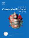
Three-dimensional reconstruction based on computed tomography/magnetic resonance imaging multimodal image fusion for parotid gland tumor diagnosis and treatment
This research evaluated a novel computed tomography (CT)/magnetic resonance imaging (MRI) fusion-based three-dimensional (3D) reconstruction method for parotid gland tumor diagnosis and treatment. In 11 patients with primary parotid tumors adjacent to the facial nerve, contrast-enhanced CT and 3D multi-echo in steady acquisition MRI images were automatically fused, segmented, and reconstructed to visualize tumors and critical anatomical structures. Preoperative tumor localization and tumor–nerve relationship assessments matched intraoperative findings in 90.9 % of cases. The method outperformed facial nerve line identification in deep lobe tumor assessment. Mean deviation distances for total, nerve, vascular, and tumor markers were 3.14 ± 1.80, 3.51 ± 1.85, 2.40 ± 1.49, and 2.47 ± 2.04 mm, respectively. Of five cases with facial nerve invasion on 3D reconstruction, three received nerve sacrifice and anastomosis, whereas two with nerve continuity preservation received postoperative radiotherapy. This technique enables precise spatial mapping of parotid tumors, aiding surgical planning and facial nerve preservation.
求助全文
通过发布文献求助,成功后即可免费获取论文全文。 去求助
来源期刊
CiteScore
5.20
自引率
22.60%
发文量
117
审稿时长
70 days
期刊介绍: The Journal of Cranio-Maxillofacial Surgery publishes articles covering all aspects of surgery of the head, face and jaw. Specific topics covered recently have included: • Distraction osteogenesis • Synthetic bone substitutes • Fibroblast growth factors • Fetal wound healing • Skull base surgery • Computer-assisted surgery • Vascularized bone grafts
×
引用
GB/T 7714-2015
复制
MLA
复制
APA
复制
导出至
BibTeX EndNote RefMan NoteFirst NoteExpress
×
提示
您的信息不完整,为了账户安全,请先补充。
现在去补充
×
提示
您因"违规操作"
具体请查看互助需知
我知道了
×
提示
确定
请完成安全验证×
copy
已复制链接
快去分享给好友吧!
我知道了
右上角分享
点击右上角分享
0
联系我们:info@booksci.cn Book学术提供免费学术资源搜索服务,方便国内外学者检索中英文文献。致力于提供最便捷和优质的服务体验。 Copyright © 2023 布克学术 All rights reserved.
京ICP备2023020795号-1
ghs 京公网安备 11010802042870号
Book学术文献互助
Book学术文献互助群
群 号:604180095
Book学术官方微信