{"title":"近端深静脉血栓形成患者血栓信号强度与肺栓塞的关系:磁共振成像研究。","authors":"Xinyu Wang, Congrui Sun, Yuehong Liu, Yichen Tang, Chen Zhang, Alto Stemmer, Jiajia Zhang, Guoxi Xie, Chunmin Li, Qi Yang","doi":"10.1161/ATVBAHA.125.322857","DOIUrl":null,"url":null,"abstract":"<p><strong>Background: </strong>Approximately 50% of deep vein thrombosis (DVT) occurrences in the lower extremity proximal veins are associated with pulmonary embolism (PE). The progression of proximal DVT is critical in PE development, as reflected by thrombus composition. Magnetic resonance black-blood thrombus imaging can identify venous thrombus components by displaying thrombus signal intensity variations. This study investigated the association between thrombus signal intensity and PE in patients with proximal DVT.</p><p><strong>Methods: </strong>A total of 126 patients with proximal DVT were recruited, and all underwent magnetic resonance black-blood thrombus imaging examination. The patients were divided into 2 groups: DVT-only and DVT with PE. The whole thrombus signal intensity ratio, proximal thrombus signal intensity ratio, distal thrombus signal intensity ratio, and thrombus volume were assessed. Histological analysis was performed to characterize the thrombus content. Logistic regression models were performed to evaluate the relationship between thrombus signal intensity and PE.</p><p><strong>Results: </strong>Of the 126 eligible patients, 73 (58%) patients were in the DVT with PE group. Both proximal thrombus signal intensity ratio and whole thrombus signal intensity ratio were lower in the DVT with PE group compared with the DVT-only group (1.92±0.54 versus 1.31±0.42, <i>P</i><0.001; 1.76±0.41 versus 1.62±0.36, <i>P</i>=0.034). The percentage of fibrin area (13.99±1.56% versus 7.51±1.25%, <i>P</i>=0.0087) and red blood cells area (49.65±18.8% versus 13.41±4.74%, <i>P</i>=0.0012) were higher in DVT with PE than DVT-only group. Univariate and multivariate logistic regression analysis showed that proximal thrombus signal intensity ratio remained statistically significant (odds ratio, 0.21 [95% CI, 0.12-0.39]; <i>P</i><0.001).</p><p><strong>Conclusions: </strong>The proximal thrombus signal intensity ratio of the thrombus was independently associated with acute PE in patients with proximal lower extremity DVT, suggesting that thrombus components may be important in PE occurrence. These findings could provide novel insights for understanding DVT evolution.</p>","PeriodicalId":8401,"journal":{"name":"Arteriosclerosis, Thrombosis, and Vascular Biology","volume":" ","pages":"1957-1968"},"PeriodicalIF":7.4000,"publicationDate":"2025-10-01","publicationTypes":"Journal Article","fieldsOfStudy":null,"isOpenAccess":false,"openAccessPdf":"https://www.ncbi.nlm.nih.gov/pmc/articles/PMC12453106/pdf/","citationCount":"0","resultStr":"{\"title\":\"Association Between Thrombus Signal Intensity and Pulmonary Embolism in Patients With Proximal Deep Vein Thrombosis: A Magnetic Resonance Imaging Study.\",\"authors\":\"Xinyu Wang, Congrui Sun, Yuehong Liu, Yichen Tang, Chen Zhang, Alto Stemmer, Jiajia Zhang, Guoxi Xie, Chunmin Li, Qi Yang\",\"doi\":\"10.1161/ATVBAHA.125.322857\",\"DOIUrl\":null,\"url\":null,\"abstract\":\"<p><strong>Background: </strong>Approximately 50% of deep vein thrombosis (DVT) occurrences in the lower extremity proximal veins are associated with pulmonary embolism (PE). The progression of proximal DVT is critical in PE development, as reflected by thrombus composition. Magnetic resonance black-blood thrombus imaging can identify venous thrombus components by displaying thrombus signal intensity variations. This study investigated the association between thrombus signal intensity and PE in patients with proximal DVT.</p><p><strong>Methods: </strong>A total of 126 patients with proximal DVT were recruited, and all underwent magnetic resonance black-blood thrombus imaging examination. The patients were divided into 2 groups: DVT-only and DVT with PE. The whole thrombus signal intensity ratio, proximal thrombus signal intensity ratio, distal thrombus signal intensity ratio, and thrombus volume were assessed. Histological analysis was performed to characterize the thrombus content. Logistic regression models were performed to evaluate the relationship between thrombus signal intensity and PE.</p><p><strong>Results: </strong>Of the 126 eligible patients, 73 (58%) patients were in the DVT with PE group. Both proximal thrombus signal intensity ratio and whole thrombus signal intensity ratio were lower in the DVT with PE group compared with the DVT-only group (1.92±0.54 versus 1.31±0.42, <i>P</i><0.001; 1.76±0.41 versus 1.62±0.36, <i>P</i>=0.034). The percentage of fibrin area (13.99±1.56% versus 7.51±1.25%, <i>P</i>=0.0087) and red blood cells area (49.65±18.8% versus 13.41±4.74%, <i>P</i>=0.0012) were higher in DVT with PE than DVT-only group. Univariate and multivariate logistic regression analysis showed that proximal thrombus signal intensity ratio remained statistically significant (odds ratio, 0.21 [95% CI, 0.12-0.39]; <i>P</i><0.001).</p><p><strong>Conclusions: </strong>The proximal thrombus signal intensity ratio of the thrombus was independently associated with acute PE in patients with proximal lower extremity DVT, suggesting that thrombus components may be important in PE occurrence. These findings could provide novel insights for understanding DVT evolution.</p>\",\"PeriodicalId\":8401,\"journal\":{\"name\":\"Arteriosclerosis, Thrombosis, and Vascular Biology\",\"volume\":\" \",\"pages\":\"1957-1968\"},\"PeriodicalIF\":7.4000,\"publicationDate\":\"2025-10-01\",\"publicationTypes\":\"Journal Article\",\"fieldsOfStudy\":null,\"isOpenAccess\":false,\"openAccessPdf\":\"https://www.ncbi.nlm.nih.gov/pmc/articles/PMC12453106/pdf/\",\"citationCount\":\"0\",\"resultStr\":null,\"platform\":\"Semanticscholar\",\"paperid\":null,\"PeriodicalName\":\"Arteriosclerosis, Thrombosis, and Vascular Biology\",\"FirstCategoryId\":\"3\",\"ListUrlMain\":\"https://doi.org/10.1161/ATVBAHA.125.322857\",\"RegionNum\":1,\"RegionCategory\":\"医学\",\"ArticlePicture\":[],\"TitleCN\":null,\"AbstractTextCN\":null,\"PMCID\":null,\"EPubDate\":\"2025/8/14 0:00:00\",\"PubModel\":\"Epub\",\"JCR\":\"Q1\",\"JCRName\":\"HEMATOLOGY\",\"Score\":null,\"Total\":0}","platform":"Semanticscholar","paperid":null,"PeriodicalName":"Arteriosclerosis, Thrombosis, and Vascular Biology","FirstCategoryId":"3","ListUrlMain":"https://doi.org/10.1161/ATVBAHA.125.322857","RegionNum":1,"RegionCategory":"医学","ArticlePicture":[],"TitleCN":null,"AbstractTextCN":null,"PMCID":null,"EPubDate":"2025/8/14 0:00:00","PubModel":"Epub","JCR":"Q1","JCRName":"HEMATOLOGY","Score":null,"Total":0}
引用次数: 0
摘要
背景:大约50%的下肢近端静脉深静脉血栓形成(DVT)与肺栓塞(PE)有关。近端DVT的进展对PE的发展至关重要,正如血栓组成所反映的那样。磁共振黑血血栓成像可以通过显示血栓信号强度的变化来识别静脉血栓成分。本研究探讨近端DVT患者血栓信号强度与PE之间的关系。方法:选取126例近端深静脉血栓患者,行磁共振黑血血栓显像检查。将患者分为单纯DVT组和DVT合并PE组。评估全血栓信号强度比、近端血栓信号强度比、远端血栓信号强度比、血栓体积。进行组织学分析以表征血栓的含量。采用Logistic回归模型评估血栓信号强度与PE之间的关系。结果:在126例符合条件的患者中,73例(58%)患者属于DVT合并PE组。DVT合并PE组近端血栓信号强度比和全端血栓信号强度比均低于单纯DVT组(1.92±0.54 vs 1.31±0.42,PP=0.034)。合并PE组纤维蛋白面积(13.99±1.56%比7.51±1.25%,P=0.0087)和红细胞面积(49.65±18.8%比13.41±4.74%,P=0.0012)均高于单纯DVT组。单因素和多因素logistic回归分析显示,近端血栓信号强度比仍具有统计学意义(优势比0.21 [95% CI, 0.12-0.39];结论:下肢近端DVT患者血栓近端信号强度比与急性PE独立相关,提示血栓成分可能在PE发生中起重要作用。这些发现可能为理解深静脉血栓的进化提供新的见解。
Association Between Thrombus Signal Intensity and Pulmonary Embolism in Patients With Proximal Deep Vein Thrombosis: A Magnetic Resonance Imaging Study.
Background: Approximately 50% of deep vein thrombosis (DVT) occurrences in the lower extremity proximal veins are associated with pulmonary embolism (PE). The progression of proximal DVT is critical in PE development, as reflected by thrombus composition. Magnetic resonance black-blood thrombus imaging can identify venous thrombus components by displaying thrombus signal intensity variations. This study investigated the association between thrombus signal intensity and PE in patients with proximal DVT.
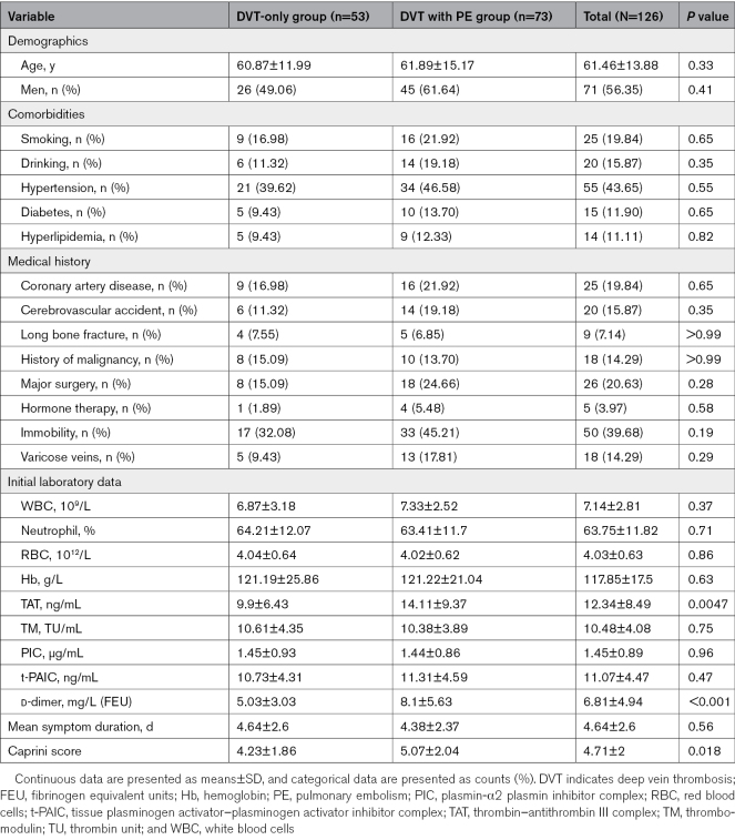
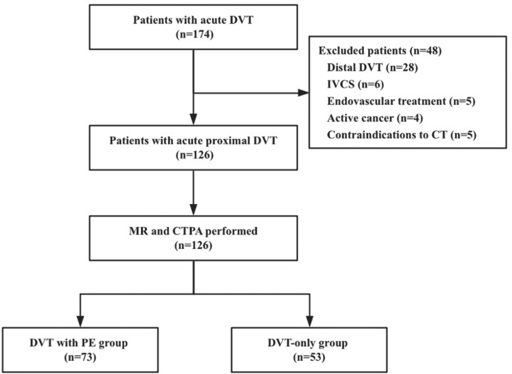
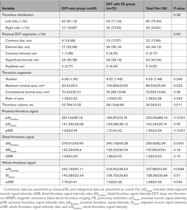
Methods: A total of 126 patients with proximal DVT were recruited, and all underwent magnetic resonance black-blood thrombus imaging examination. The patients were divided into 2 groups: DVT-only and DVT with PE. The whole thrombus signal intensity ratio, proximal thrombus signal intensity ratio, distal thrombus signal intensity ratio, and thrombus volume were assessed. Histological analysis was performed to characterize the thrombus content. Logistic regression models were performed to evaluate the relationship between thrombus signal intensity and PE.
Results: Of the 126 eligible patients, 73 (58%) patients were in the DVT with PE group. Both proximal thrombus signal intensity ratio and whole thrombus signal intensity ratio were lower in the DVT with PE group compared with the DVT-only group (1.92±0.54 versus 1.31±0.42, P<0.001; 1.76±0.41 versus 1.62±0.36, P=0.034). The percentage of fibrin area (13.99±1.56% versus 7.51±1.25%, P=0.0087) and red blood cells area (49.65±18.8% versus 13.41±4.74%, P=0.0012) were higher in DVT with PE than DVT-only group. Univariate and multivariate logistic regression analysis showed that proximal thrombus signal intensity ratio remained statistically significant (odds ratio, 0.21 [95% CI, 0.12-0.39]; P<0.001).
Conclusions: The proximal thrombus signal intensity ratio of the thrombus was independently associated with acute PE in patients with proximal lower extremity DVT, suggesting that thrombus components may be important in PE occurrence. These findings could provide novel insights for understanding DVT evolution.
期刊介绍:
The journal "Arteriosclerosis, Thrombosis, and Vascular Biology" (ATVB) is a scientific publication that focuses on the fields of vascular biology, atherosclerosis, and thrombosis. It is a peer-reviewed journal that publishes original research articles, reviews, and other scholarly content related to these areas. The journal is published by the American Heart Association (AHA) and the American Stroke Association (ASA).
The journal was published bi-monthly until January 1992, after which it transitioned to a monthly publication schedule. The journal is aimed at a professional audience, including academic cardiologists, vascular biologists, physiologists, pharmacologists and hematologists.
 求助内容:
求助内容: 应助结果提醒方式:


