皮肤结外NK/T细胞淋巴瘤:根据原发肿瘤部位的空间转录组谱异质性
IF 5
2区 医学
Q2 Medicine
引用次数: 0
摘要
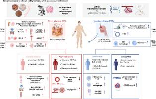
结外NK/ t细胞淋巴瘤(ENKTL)伴皮肤受累可分为原发性皮肤ENKTL (pcENKTL)和继发性皮肤受累(scENKTL)。尽管起源不同,临床和组织病理学的发现是难以区分的。此外,每个实体的预后决定因素尚未确定。我们研究了pcENKTL和scENKTL在空间分辨转录组谱上的差异。材料和方法采用CD56和CD3形态学标记,选择7个样本,共24个感兴趣区域进行pcENKTCL和scENKTL的检测。结果在cd56阳性肿瘤细胞区,与scENKTL相比,pcENKTL检测到91个差异表达基因(DEGs)上调,27个差异表达基因(DEGs)下调。蛋白-蛋白相互作用网络揭示了pcENKTL中干扰素信号、T细胞受体信号和程序性细胞死亡的显著富集。此外,在scENKTL中观察到翻译途径的显著富集和无义介导的衰变。在免疫细胞区,pcENKTL中骨髓树突状细胞(p = 0.044)和M1巨噬细胞(p = 0.039)数量增加。相反,scENKTL的中性粒细胞(p = 0.030)和M2巨噬细胞(p = 0.030)数量增加。我们发现完全缓解的pcENKTL患者免疫应答和抗原呈递增加,而疾病进展的pcENKTL患者血管生成增加。另外,长生存期的scENKTL表现出HLA表达和CD8记忆T细胞和M1巨噬细胞的增加,而短生存期的scENKTL表现出癌症相关成纤维细胞和BIRC5的增加。结论总的来说,pcENKTL和scENKTL在转录组表达、肿瘤微环境以及基于预后的亚群上的差异可以拓宽我们对ENKTL生物学特性的认识。本文章由计算机程序翻译,如有差异,请以英文原文为准。
Cutaneous extranodal NK/T cell lymphoma: heterogeneity of spatial transcriptomic profiling according to primary tumor site
Introduction
Extranodal NK/T-cell lymphoma (ENKTL) with cutaneous involvement can be divided into primary cutaneous ENKTL (pcENKTL) and secondary cutaneous involvement (scENKTL). Despite different originations, the clinical and histopathological findings are indistinguishable. Moreover, the prognosis determinants in each entity have yet to be established. We investigated differences in spatially resolved transcriptome profiles between pcENKTL and scENKTL.
Materials and methods
Seven samples with 24 regions of interest were selected for pcENKTCL and scENKTL, using CD56 and CD3 morphology markers.
Results
In CD56-positive tumor cell areas, we detected 91 upregulated differentially expressed genes (DEGs) and 27 downregulated DEGs in pcENKTL compared with scENKTL. Protein–protein interaction network revealed significant enrichment of interferon signaling, T cell receptor signaling, and programmed cell death in pcENKTL. Moreover, significant enrichment of translation pathways and nonsense-mediated decay in scENKTL was observed. In immune cell areas, myeloid dendritic cell (p = 0.044) and M1 macrophage (p = 0.039) numbers were increased in pcENKTL. Conversely, neutrophil (p = 0.030) and M2 macrophage (p = 0.030) numbers were increased in scENKTL. We found an increased immune response and antigen presentation in pcENKTL with complete remission, while pcENKTL with progressive disease showed increased angiogenesis. Alternatively, scENKTL with long survival showed increased HLA expression and CD8 memory T cells and M1 macrophages, while scENKTL with short survival showed increased cancer-associated fibroblasts and BIRC5.
Conclusion
Overall, the differences in transcriptomic expression and tumor microenvironment between pcENKTL and scENKTL, as well as subgroups based on the prognosis could widen our understanding of the biological characteristics of ENKTL.
求助全文
通过发布文献求助,成功后即可免费获取论文全文。
去求助
来源期刊
Translational Oncology
ONCOLOGY-
CiteScore
8.40
自引率
2.00%
发文量
314
审稿时长
54 days
期刊介绍:
Translational Oncology publishes the results of novel research investigations which bridge the laboratory and clinical settings including risk assessment, cellular and molecular characterization, prevention, detection, diagnosis and treatment of human cancers with the overall goal of improving the clinical care of oncology patients. Translational Oncology will publish laboratory studies of novel therapeutic interventions as well as clinical trials which evaluate new treatment paradigms for cancer. Peer reviewed manuscript types include Original Reports, Reviews and Editorials.
 求助内容:
求助内容: 应助结果提醒方式:


