{"title":"骨报告和CT数据系统(Bone- rads -CT):四名读者对来自三个本地和两个公共数据库的328例病例的验证研究。","authors":"Yue Xing, Defang Ding, Shun Dai, Yangfan Hu, Xianwei Liu, Liangjing Lyu, Guangcheng Zhang, Shiqi Mao, Qian Yin, Junjie Lu, Jiarui Yang, Yang Song, Huan Zhang, Chengzhou Li, Weiwu Yao, Jingyu Zhong","doi":"10.1186/s13244-025-02057-8","DOIUrl":null,"url":null,"abstract":"<p><strong>Objective: </strong>To evaluate the reproducibility and effectiveness of the bone reporting and data system on CT (Bone-RADS-CT) for incidental solitary bone lesions in adults.</p><p><strong>Materials and methods: </strong>We retrospectively included 328 CT cases from three local and two public databases, respectively. All the cases were histopathologically or clinically confirmed bone lesions, \"do not touch\" lesions with typical appearance, and remained stable for at least 2 years. Each lesion with gender, age, and clinical history was categorized according to the Bone-RADS algorithm by two musculoskeletal radiologists and two non-musculoskeletal radiologists. The Bone-RADS categories were as follows: Bone-RADS-1, likely benign, leave alone; Bone-RADS-2, incomplete assessed on imaging, perform different imaging modality; Bone-RADS-3, intermediate, perform follow-up imaging; Bone-RADS-4, suspicious for malignancy or need for treatment, biopsy and/or oncologic referral. Inter-reader agreement was evaluated. The diagnostic performance of the Bone-RADS-CT for distinguishing positive cases (intermediate or malignant lesions or osteomyelitis) from negative cases (benign lesions), were measured, using histopathology results, clinical diagnosis, or follow-up as a standard reference.</p><p><strong>Results: </strong>There were 223 positive cases and 105 negative cases, respectively. The overall inter-reader agreement between two musculoskeletal and two non-musculoskeletal radiologists were both moderate (weighted kappa 0.553 and 0.403). The diagnostic performance for identifying intermediate or malignant lesions or osteomyelitis ranged according to radiologists with sensitivities of 88.8% to 94.6%, specificities of 42.9% to 71.1%, and accuracies of 78.0% to 86.6%.</p><p><strong>Conclusion: </strong>Bone-RADS-CT is effective for identifying bone lesions that need further treatment, but is only moderately reliable for readers of different specialties and experience.</p><p><strong>Critical relevance statement: </strong>Bone-RADS-CT has been demonstrated to be a reliable algorithm for non-musculoskeletal radiologists and an effective tool for identifying the \"need for treatment\" incidental solitary bone lesions in adults, but still needs improvement in the rating method and category definition.</p><p><strong>Key points: </strong>Bone-RADS-CT has been demonstrated to be reliable and accurate when rated by musculoskeletal radiologists. Bone-RADS-CT achieved moderate agreement for musculoskeletal and non-musculoskeletal radiologists. Bone-RADS-CT presented high sensitivities but low specificities for identifying \"need for treatment\" bone lesions.</p>","PeriodicalId":13639,"journal":{"name":"Insights into Imaging","volume":"16 1","pages":"174"},"PeriodicalIF":4.5000,"publicationDate":"2025-08-12","publicationTypes":"Journal Article","fieldsOfStudy":null,"isOpenAccess":false,"openAccessPdf":"https://www.ncbi.nlm.nih.gov/pmc/articles/PMC12344047/pdf/","citationCount":"0","resultStr":"{\"title\":\"Bone reporting and data system on CT (Bone-RADS-CT): a validation study by four readers on 328 cases from three local and two public databases.\",\"authors\":\"Yue Xing, Defang Ding, Shun Dai, Yangfan Hu, Xianwei Liu, Liangjing Lyu, Guangcheng Zhang, Shiqi Mao, Qian Yin, Junjie Lu, Jiarui Yang, Yang Song, Huan Zhang, Chengzhou Li, Weiwu Yao, Jingyu Zhong\",\"doi\":\"10.1186/s13244-025-02057-8\",\"DOIUrl\":null,\"url\":null,\"abstract\":\"<p><strong>Objective: </strong>To evaluate the reproducibility and effectiveness of the bone reporting and data system on CT (Bone-RADS-CT) for incidental solitary bone lesions in adults.</p><p><strong>Materials and methods: </strong>We retrospectively included 328 CT cases from three local and two public databases, respectively. All the cases were histopathologically or clinically confirmed bone lesions, \\\"do not touch\\\" lesions with typical appearance, and remained stable for at least 2 years. Each lesion with gender, age, and clinical history was categorized according to the Bone-RADS algorithm by two musculoskeletal radiologists and two non-musculoskeletal radiologists. The Bone-RADS categories were as follows: Bone-RADS-1, likely benign, leave alone; Bone-RADS-2, incomplete assessed on imaging, perform different imaging modality; Bone-RADS-3, intermediate, perform follow-up imaging; Bone-RADS-4, suspicious for malignancy or need for treatment, biopsy and/or oncologic referral. Inter-reader agreement was evaluated. The diagnostic performance of the Bone-RADS-CT for distinguishing positive cases (intermediate or malignant lesions or osteomyelitis) from negative cases (benign lesions), were measured, using histopathology results, clinical diagnosis, or follow-up as a standard reference.</p><p><strong>Results: </strong>There were 223 positive cases and 105 negative cases, respectively. The overall inter-reader agreement between two musculoskeletal and two non-musculoskeletal radiologists were both moderate (weighted kappa 0.553 and 0.403). The diagnostic performance for identifying intermediate or malignant lesions or osteomyelitis ranged according to radiologists with sensitivities of 88.8% to 94.6%, specificities of 42.9% to 71.1%, and accuracies of 78.0% to 86.6%.</p><p><strong>Conclusion: </strong>Bone-RADS-CT is effective for identifying bone lesions that need further treatment, but is only moderately reliable for readers of different specialties and experience.</p><p><strong>Critical relevance statement: </strong>Bone-RADS-CT has been demonstrated to be a reliable algorithm for non-musculoskeletal radiologists and an effective tool for identifying the \\\"need for treatment\\\" incidental solitary bone lesions in adults, but still needs improvement in the rating method and category definition.</p><p><strong>Key points: </strong>Bone-RADS-CT has been demonstrated to be reliable and accurate when rated by musculoskeletal radiologists. Bone-RADS-CT achieved moderate agreement for musculoskeletal and non-musculoskeletal radiologists. Bone-RADS-CT presented high sensitivities but low specificities for identifying \\\"need for treatment\\\" bone lesions.</p>\",\"PeriodicalId\":13639,\"journal\":{\"name\":\"Insights into Imaging\",\"volume\":\"16 1\",\"pages\":\"174\"},\"PeriodicalIF\":4.5000,\"publicationDate\":\"2025-08-12\",\"publicationTypes\":\"Journal Article\",\"fieldsOfStudy\":null,\"isOpenAccess\":false,\"openAccessPdf\":\"https://www.ncbi.nlm.nih.gov/pmc/articles/PMC12344047/pdf/\",\"citationCount\":\"0\",\"resultStr\":null,\"platform\":\"Semanticscholar\",\"paperid\":null,\"PeriodicalName\":\"Insights into Imaging\",\"FirstCategoryId\":\"3\",\"ListUrlMain\":\"https://doi.org/10.1186/s13244-025-02057-8\",\"RegionNum\":2,\"RegionCategory\":\"医学\",\"ArticlePicture\":[],\"TitleCN\":null,\"AbstractTextCN\":null,\"PMCID\":null,\"EPubDate\":\"\",\"PubModel\":\"\",\"JCR\":\"Q1\",\"JCRName\":\"RADIOLOGY, NUCLEAR MEDICINE & MEDICAL IMAGING\",\"Score\":null,\"Total\":0}","platform":"Semanticscholar","paperid":null,"PeriodicalName":"Insights into Imaging","FirstCategoryId":"3","ListUrlMain":"https://doi.org/10.1186/s13244-025-02057-8","RegionNum":2,"RegionCategory":"医学","ArticlePicture":[],"TitleCN":null,"AbstractTextCN":null,"PMCID":null,"EPubDate":"","PubModel":"","JCR":"Q1","JCRName":"RADIOLOGY, NUCLEAR MEDICINE & MEDICAL IMAGING","Score":null,"Total":0}
Bone reporting and data system on CT (Bone-RADS-CT): a validation study by four readers on 328 cases from three local and two public databases.
Objective: To evaluate the reproducibility and effectiveness of the bone reporting and data system on CT (Bone-RADS-CT) for incidental solitary bone lesions in adults.
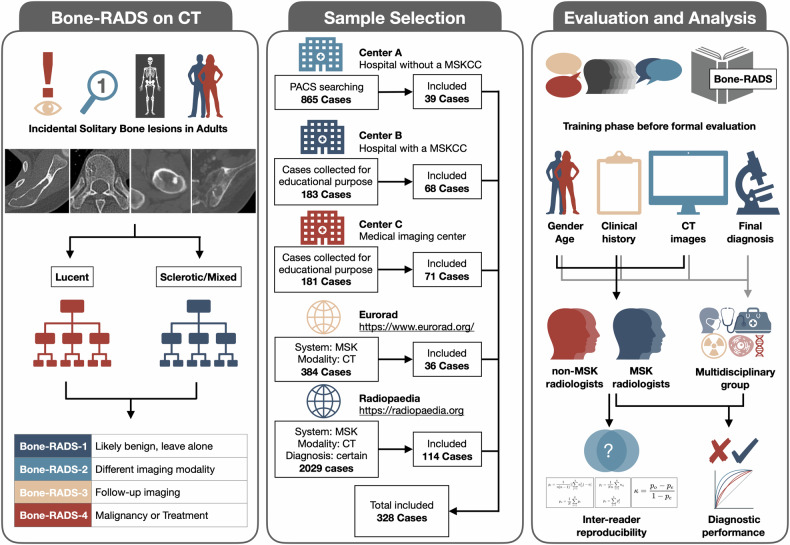
Materials and methods: We retrospectively included 328 CT cases from three local and two public databases, respectively. All the cases were histopathologically or clinically confirmed bone lesions, "do not touch" lesions with typical appearance, and remained stable for at least 2 years. Each lesion with gender, age, and clinical history was categorized according to the Bone-RADS algorithm by two musculoskeletal radiologists and two non-musculoskeletal radiologists. The Bone-RADS categories were as follows: Bone-RADS-1, likely benign, leave alone; Bone-RADS-2, incomplete assessed on imaging, perform different imaging modality; Bone-RADS-3, intermediate, perform follow-up imaging; Bone-RADS-4, suspicious for malignancy or need for treatment, biopsy and/or oncologic referral. Inter-reader agreement was evaluated. The diagnostic performance of the Bone-RADS-CT for distinguishing positive cases (intermediate or malignant lesions or osteomyelitis) from negative cases (benign lesions), were measured, using histopathology results, clinical diagnosis, or follow-up as a standard reference.
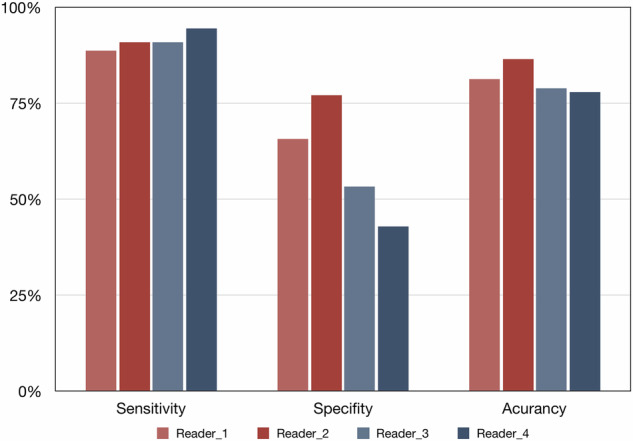
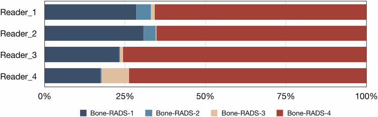
Results: There were 223 positive cases and 105 negative cases, respectively. The overall inter-reader agreement between two musculoskeletal and two non-musculoskeletal radiologists were both moderate (weighted kappa 0.553 and 0.403). The diagnostic performance for identifying intermediate or malignant lesions or osteomyelitis ranged according to radiologists with sensitivities of 88.8% to 94.6%, specificities of 42.9% to 71.1%, and accuracies of 78.0% to 86.6%.
Conclusion: Bone-RADS-CT is effective for identifying bone lesions that need further treatment, but is only moderately reliable for readers of different specialties and experience.
Critical relevance statement: Bone-RADS-CT has been demonstrated to be a reliable algorithm for non-musculoskeletal radiologists and an effective tool for identifying the "need for treatment" incidental solitary bone lesions in adults, but still needs improvement in the rating method and category definition.
Key points: Bone-RADS-CT has been demonstrated to be reliable and accurate when rated by musculoskeletal radiologists. Bone-RADS-CT achieved moderate agreement for musculoskeletal and non-musculoskeletal radiologists. Bone-RADS-CT presented high sensitivities but low specificities for identifying "need for treatment" bone lesions.
期刊介绍:
Insights into Imaging (I³) is a peer-reviewed open access journal published under the brand SpringerOpen. All content published in the journal is freely available online to anyone, anywhere!
I³ continuously updates scientific knowledge and progress in best-practice standards in radiology through the publication of original articles and state-of-the-art reviews and opinions, along with recommendations and statements from the leading radiological societies in Europe.
Founded by the European Society of Radiology (ESR), I³ creates a platform for educational material, guidelines and recommendations, and a forum for topics of controversy.
A balanced combination of review articles, original papers, short communications from European radiological congresses and information on society matters makes I³ an indispensable source for current information in this field.
I³ is owned by the ESR, however authors retain copyright to their article according to the Creative Commons Attribution License (see Copyright and License Agreement). All articles can be read, redistributed and reused for free, as long as the author of the original work is cited properly.
The open access fees (article-processing charges) for this journal are kindly sponsored by ESR for all Members.
The journal went open access in 2012, which means that all articles published since then are freely available online.

 求助内容:
求助内容: 应助结果提醒方式:


