Kamil Balaban, Ramazan Akmeşe, Hüseyin Hakan Kınık, Mahmut Kalem
{"title":"胫距跟骨关节融合术伴逆行髓内钉:入路改变有何不同?","authors":"Kamil Balaban, Ramazan Akmeşe, Hüseyin Hakan Kınık, Mahmut Kalem","doi":"10.52312/jdrs.2025.2380","DOIUrl":null,"url":null,"abstract":"<p><strong>Objectives: </strong>This study aims to compare the clinical and radiographic outcomes of open (lateral transfibular) and arthroscopic joint debridement techniques in tibiotalocalcaneal arthrodesis (TTCA) using the same nail system.</p><p><strong>Patients and methods: </strong>Between January 2011 and December 2022, a total of 62 patients (21 males, 41 females; mean age 53.81±16.68 years; range 18 to 82 years) who underwent TTCA with retrograde intramedullary nail were retrospectively analyzed. The patients were classified as open (n=30) or arthroscopy (n=32) based on the method used for joint debridement. Data including demographic characteristics, pre-and postoperative radiographs, skin-to-skin operative times, and fluoroscopy times were recorded. Tibiotalar and subtalar union rates, coronal and sagittal ankle alignment examined through coronal tibiotalar (CTT) and sagittal tibiotalar (STT) angles were also noted. Functional outcomes were measured using the American Orthopaedic Foot and Ankle Society Ankle-Hindfoot Score (AOFAS-AHS) and Visual Analog Scale (VAS). Complications were evaluated.</p><p><strong>Results: </strong>A total of 34 ankles (n=30) underwent open TTCA, while 34 ankles (n=32) had arthroscopic TTCA. Baseline characteristics and follow-up duration were similar between the groups (p>0.05). The overall fusion rate (tibiotalar and subtalar) was 94.1% in the open group and 85.3% in the arthroscopic group (p=0.425). Both open and arthroscopy groups achieved satisfactory coronal and sagittal ankle alignment. The median CTT angles were 94° and 91°, and STT angles were 109° and 112°, respectively. The arthroscopy group had significantly shorter operative time, fluoroscopy time, and hospital stay (p<0.001, p=0.019, p<0.001, respectively). No significant differences were found in complication rates, postoperative AOFAS-AHS, and VAS scores (p>0.05).</p><p><strong>Conclusion: </strong>Both open and arthroscopic TTCA approaches yielded similar radiographic and clinical outcomes. Based on these findings, we can speculate that the arthroscopic technique may offer advantages in perioperative efficiency, suggesting it is a viable alternative in appropriately selected patients.</p>","PeriodicalId":73560,"journal":{"name":"Joint diseases and related surgery","volume":"36 3","pages":"711-723"},"PeriodicalIF":1.9000,"publicationDate":"2025-06-27","publicationTypes":"Journal Article","fieldsOfStudy":null,"isOpenAccess":false,"openAccessPdf":"https://www.ncbi.nlm.nih.gov/pmc/articles/PMC12456346/pdf/","citationCount":"0","resultStr":"{\"title\":\"Tibiotalocalcaneal arthrodesis with retrograde intramedullary nailing: What differs only approach change?\",\"authors\":\"Kamil Balaban, Ramazan Akmeşe, Hüseyin Hakan Kınık, Mahmut Kalem\",\"doi\":\"10.52312/jdrs.2025.2380\",\"DOIUrl\":null,\"url\":null,\"abstract\":\"<p><strong>Objectives: </strong>This study aims to compare the clinical and radiographic outcomes of open (lateral transfibular) and arthroscopic joint debridement techniques in tibiotalocalcaneal arthrodesis (TTCA) using the same nail system.</p><p><strong>Patients and methods: </strong>Between January 2011 and December 2022, a total of 62 patients (21 males, 41 females; mean age 53.81±16.68 years; range 18 to 82 years) who underwent TTCA with retrograde intramedullary nail were retrospectively analyzed. The patients were classified as open (n=30) or arthroscopy (n=32) based on the method used for joint debridement. Data including demographic characteristics, pre-and postoperative radiographs, skin-to-skin operative times, and fluoroscopy times were recorded. Tibiotalar and subtalar union rates, coronal and sagittal ankle alignment examined through coronal tibiotalar (CTT) and sagittal tibiotalar (STT) angles were also noted. Functional outcomes were measured using the American Orthopaedic Foot and Ankle Society Ankle-Hindfoot Score (AOFAS-AHS) and Visual Analog Scale (VAS). Complications were evaluated.</p><p><strong>Results: </strong>A total of 34 ankles (n=30) underwent open TTCA, while 34 ankles (n=32) had arthroscopic TTCA. Baseline characteristics and follow-up duration were similar between the groups (p>0.05). The overall fusion rate (tibiotalar and subtalar) was 94.1% in the open group and 85.3% in the arthroscopic group (p=0.425). Both open and arthroscopy groups achieved satisfactory coronal and sagittal ankle alignment. The median CTT angles were 94° and 91°, and STT angles were 109° and 112°, respectively. The arthroscopy group had significantly shorter operative time, fluoroscopy time, and hospital stay (p<0.001, p=0.019, p<0.001, respectively). No significant differences were found in complication rates, postoperative AOFAS-AHS, and VAS scores (p>0.05).</p><p><strong>Conclusion: </strong>Both open and arthroscopic TTCA approaches yielded similar radiographic and clinical outcomes. Based on these findings, we can speculate that the arthroscopic technique may offer advantages in perioperative efficiency, suggesting it is a viable alternative in appropriately selected patients.</p>\",\"PeriodicalId\":73560,\"journal\":{\"name\":\"Joint diseases and related surgery\",\"volume\":\"36 3\",\"pages\":\"711-723\"},\"PeriodicalIF\":1.9000,\"publicationDate\":\"2025-06-27\",\"publicationTypes\":\"Journal Article\",\"fieldsOfStudy\":null,\"isOpenAccess\":false,\"openAccessPdf\":\"https://www.ncbi.nlm.nih.gov/pmc/articles/PMC12456346/pdf/\",\"citationCount\":\"0\",\"resultStr\":null,\"platform\":\"Semanticscholar\",\"paperid\":null,\"PeriodicalName\":\"Joint diseases and related surgery\",\"FirstCategoryId\":\"1085\",\"ListUrlMain\":\"https://doi.org/10.52312/jdrs.2025.2380\",\"RegionNum\":0,\"RegionCategory\":null,\"ArticlePicture\":[],\"TitleCN\":null,\"AbstractTextCN\":null,\"PMCID\":null,\"EPubDate\":\"\",\"PubModel\":\"\",\"JCR\":\"Q2\",\"JCRName\":\"ORTHOPEDICS\",\"Score\":null,\"Total\":0}","platform":"Semanticscholar","paperid":null,"PeriodicalName":"Joint diseases and related surgery","FirstCategoryId":"1085","ListUrlMain":"https://doi.org/10.52312/jdrs.2025.2380","RegionNum":0,"RegionCategory":null,"ArticlePicture":[],"TitleCN":null,"AbstractTextCN":null,"PMCID":null,"EPubDate":"","PubModel":"","JCR":"Q2","JCRName":"ORTHOPEDICS","Score":null,"Total":0}
Tibiotalocalcaneal arthrodesis with retrograde intramedullary nailing: What differs only approach change?
Objectives: This study aims to compare the clinical and radiographic outcomes of open (lateral transfibular) and arthroscopic joint debridement techniques in tibiotalocalcaneal arthrodesis (TTCA) using the same nail system.
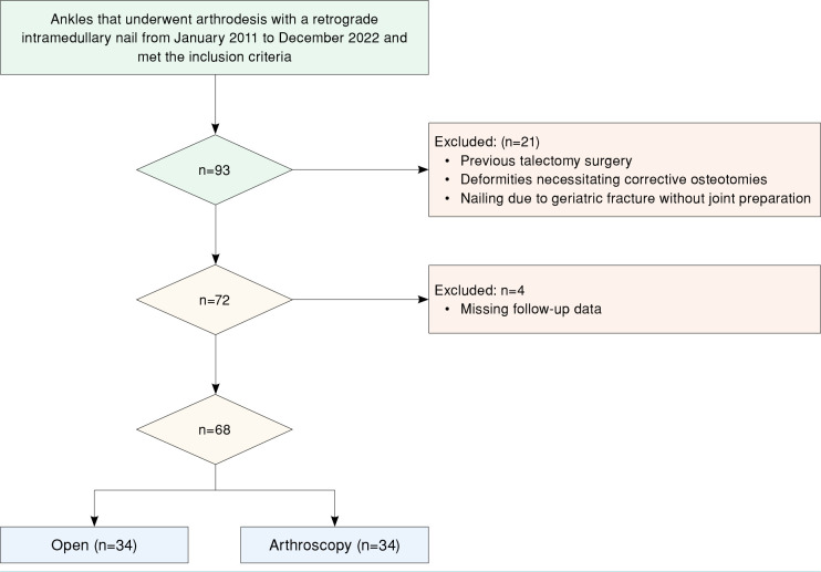
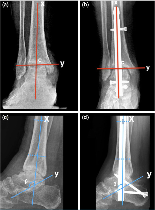
Patients and methods: Between January 2011 and December 2022, a total of 62 patients (21 males, 41 females; mean age 53.81±16.68 years; range 18 to 82 years) who underwent TTCA with retrograde intramedullary nail were retrospectively analyzed. The patients were classified as open (n=30) or arthroscopy (n=32) based on the method used for joint debridement. Data including demographic characteristics, pre-and postoperative radiographs, skin-to-skin operative times, and fluoroscopy times were recorded. Tibiotalar and subtalar union rates, coronal and sagittal ankle alignment examined through coronal tibiotalar (CTT) and sagittal tibiotalar (STT) angles were also noted. Functional outcomes were measured using the American Orthopaedic Foot and Ankle Society Ankle-Hindfoot Score (AOFAS-AHS) and Visual Analog Scale (VAS). Complications were evaluated.
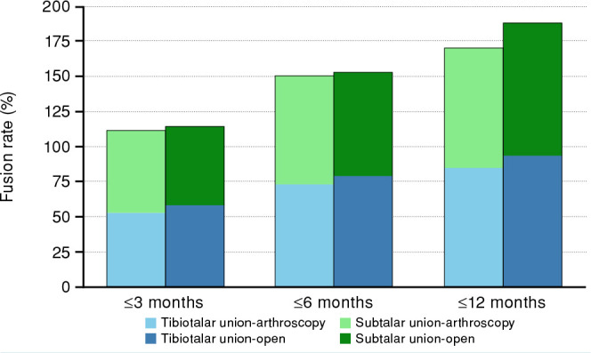
Results: A total of 34 ankles (n=30) underwent open TTCA, while 34 ankles (n=32) had arthroscopic TTCA. Baseline characteristics and follow-up duration were similar between the groups (p>0.05). The overall fusion rate (tibiotalar and subtalar) was 94.1% in the open group and 85.3% in the arthroscopic group (p=0.425). Both open and arthroscopy groups achieved satisfactory coronal and sagittal ankle alignment. The median CTT angles were 94° and 91°, and STT angles were 109° and 112°, respectively. The arthroscopy group had significantly shorter operative time, fluoroscopy time, and hospital stay (p<0.001, p=0.019, p<0.001, respectively). No significant differences were found in complication rates, postoperative AOFAS-AHS, and VAS scores (p>0.05).
Conclusion: Both open and arthroscopic TTCA approaches yielded similar radiographic and clinical outcomes. Based on these findings, we can speculate that the arthroscopic technique may offer advantages in perioperative efficiency, suggesting it is a viable alternative in appropriately selected patients.

 求助内容:
求助内容: 应助结果提醒方式:


