{"title":"心包脂肪特定成分对冠状动脉高危斑块预测的影响。","authors":"Lihong Nan, Tongli Li, Wenyu Ding, Mengshan Wu, Jintang Feng, Tianzhu Zhang, Fan Yang, Dong Li, Chunyang Sun, Ningnannan Zhang, Zhang Zhang","doi":"10.21037/qims-24-1140","DOIUrl":null,"url":null,"abstract":"<p><strong>Background: </strong>Coronary computed tomography angiography (CCTA) can be used to investigate the relationship between white adipose tissue (WAT) and brown adipose tissue (BAT) in pericardial fat (PF) and high-risk plaques (HRP) in patients with coronary artery disease (CAD). This study aimed to explore the association between specific components of PF and HRP/culprit ischemic plaques, as well as their mediating role in overall metabolic status, providing new imaging evidence for predicting adverse events in CAD.</p><p><strong>Methods: </strong>The clinical risk factors and imaging images of 107 CAD patients were retrospectively analyzed. Quantification of coronary artery calcium scores (CACS), PF, WAT, BAT, and pericardial fat attenuation (PFatt) were performed on non-contrast CT images. CCTA was used to evaluate myocardial ischemia and the presence of HRP. Fractional flow reserve derived from CCTA (CT-FFR) was performed in three major coronary arteries, with a threshold of ≤0.80 considered indicative of the presence of lesion-specific ischemia. HRP was defined as the presence of at least two of the four HRP features including positive remodeling (PR), low attenuation, napkin-ring sign (NRS), and spotty calcification. Mediator analysis was performed using Hayes (2018) Model-4.</p><p><strong>Results: </strong>A total of 107 CAD patients aged 65±8 years were included in this study. There were 49 patients (45.79%) with HRP and 57 patients (53.27%) with lesion-specific ischemia (CT-FFR ≤0.80). PF including WAT and BAT in the HRP group was significantly higher than that in the non-HRP group (PF: 241.28 <i>vs.</i> 204.94 cm<sup>3</sup>, P=0.005, WAT: 91.78 <i>vs.</i> 78.40 cm<sup>3</sup>, P=0.002, BAT: 56.25 <i>vs.</i> 46.71 cm<sup>3</sup>, P=0.008). Adding WAT to meaningful clinical risk factors and CACS increases the area under the curve (AUC) for HRP prediction {AUC [95% confidence interval (95% CI)]: 0.789 (0.692-0.887) <i>vs.</i> 0.655 (0.535-0.775), P<0.05}. Interestingly, adding PF to clinical risk factors and CACS decreased the AUC for the prediction of lesion-specific ischemia with no significant difference (P=0.083) [AUC (95% CI): 0.705 (0.592-0.817) <i>vs.</i> 0.796 (0.696-0.897), P<0.05]. Additionally, the mediation effect of PF accounted for 95.19% of the total effect of clinical risk factors on HRP (P<0.05).</p><p><strong>Conclusions: </strong>PF is associated with HRP, and clinical risk factors increase the risk of plaque rupture by promoting PF volume accumulation, especially WAT, which may be a potential predictor of HRP.</p>","PeriodicalId":54267,"journal":{"name":"Quantitative Imaging in Medicine and Surgery","volume":"15 8","pages":"7309-7322"},"PeriodicalIF":2.3000,"publicationDate":"2025-08-01","publicationTypes":"Journal Article","fieldsOfStudy":null,"isOpenAccess":false,"openAccessPdf":"https://www.ncbi.nlm.nih.gov/pmc/articles/PMC12332739/pdf/","citationCount":"0","resultStr":"{\"title\":\"Influence of specific components of pericardial fat on coronary high-risk plaque prediction.\",\"authors\":\"Lihong Nan, Tongli Li, Wenyu Ding, Mengshan Wu, Jintang Feng, Tianzhu Zhang, Fan Yang, Dong Li, Chunyang Sun, Ningnannan Zhang, Zhang Zhang\",\"doi\":\"10.21037/qims-24-1140\",\"DOIUrl\":null,\"url\":null,\"abstract\":\"<p><strong>Background: </strong>Coronary computed tomography angiography (CCTA) can be used to investigate the relationship between white adipose tissue (WAT) and brown adipose tissue (BAT) in pericardial fat (PF) and high-risk plaques (HRP) in patients with coronary artery disease (CAD). This study aimed to explore the association between specific components of PF and HRP/culprit ischemic plaques, as well as their mediating role in overall metabolic status, providing new imaging evidence for predicting adverse events in CAD.</p><p><strong>Methods: </strong>The clinical risk factors and imaging images of 107 CAD patients were retrospectively analyzed. Quantification of coronary artery calcium scores (CACS), PF, WAT, BAT, and pericardial fat attenuation (PFatt) were performed on non-contrast CT images. CCTA was used to evaluate myocardial ischemia and the presence of HRP. Fractional flow reserve derived from CCTA (CT-FFR) was performed in three major coronary arteries, with a threshold of ≤0.80 considered indicative of the presence of lesion-specific ischemia. HRP was defined as the presence of at least two of the four HRP features including positive remodeling (PR), low attenuation, napkin-ring sign (NRS), and spotty calcification. Mediator analysis was performed using Hayes (2018) Model-4.</p><p><strong>Results: </strong>A total of 107 CAD patients aged 65±8 years were included in this study. There were 49 patients (45.79%) with HRP and 57 patients (53.27%) with lesion-specific ischemia (CT-FFR ≤0.80). PF including WAT and BAT in the HRP group was significantly higher than that in the non-HRP group (PF: 241.28 <i>vs.</i> 204.94 cm<sup>3</sup>, P=0.005, WAT: 91.78 <i>vs.</i> 78.40 cm<sup>3</sup>, P=0.002, BAT: 56.25 <i>vs.</i> 46.71 cm<sup>3</sup>, P=0.008). Adding WAT to meaningful clinical risk factors and CACS increases the area under the curve (AUC) for HRP prediction {AUC [95% confidence interval (95% CI)]: 0.789 (0.692-0.887) <i>vs.</i> 0.655 (0.535-0.775), P<0.05}. Interestingly, adding PF to clinical risk factors and CACS decreased the AUC for the prediction of lesion-specific ischemia with no significant difference (P=0.083) [AUC (95% CI): 0.705 (0.592-0.817) <i>vs.</i> 0.796 (0.696-0.897), P<0.05]. Additionally, the mediation effect of PF accounted for 95.19% of the total effect of clinical risk factors on HRP (P<0.05).</p><p><strong>Conclusions: </strong>PF is associated with HRP, and clinical risk factors increase the risk of plaque rupture by promoting PF volume accumulation, especially WAT, which may be a potential predictor of HRP.</p>\",\"PeriodicalId\":54267,\"journal\":{\"name\":\"Quantitative Imaging in Medicine and Surgery\",\"volume\":\"15 8\",\"pages\":\"7309-7322\"},\"PeriodicalIF\":2.3000,\"publicationDate\":\"2025-08-01\",\"publicationTypes\":\"Journal Article\",\"fieldsOfStudy\":null,\"isOpenAccess\":false,\"openAccessPdf\":\"https://www.ncbi.nlm.nih.gov/pmc/articles/PMC12332739/pdf/\",\"citationCount\":\"0\",\"resultStr\":null,\"platform\":\"Semanticscholar\",\"paperid\":null,\"PeriodicalName\":\"Quantitative Imaging in Medicine and Surgery\",\"FirstCategoryId\":\"3\",\"ListUrlMain\":\"https://doi.org/10.21037/qims-24-1140\",\"RegionNum\":2,\"RegionCategory\":\"医学\",\"ArticlePicture\":[],\"TitleCN\":null,\"AbstractTextCN\":null,\"PMCID\":null,\"EPubDate\":\"2025/7/29 0:00:00\",\"PubModel\":\"Epub\",\"JCR\":\"Q2\",\"JCRName\":\"RADIOLOGY, NUCLEAR MEDICINE & MEDICAL IMAGING\",\"Score\":null,\"Total\":0}","platform":"Semanticscholar","paperid":null,"PeriodicalName":"Quantitative Imaging in Medicine and Surgery","FirstCategoryId":"3","ListUrlMain":"https://doi.org/10.21037/qims-24-1140","RegionNum":2,"RegionCategory":"医学","ArticlePicture":[],"TitleCN":null,"AbstractTextCN":null,"PMCID":null,"EPubDate":"2025/7/29 0:00:00","PubModel":"Epub","JCR":"Q2","JCRName":"RADIOLOGY, NUCLEAR MEDICINE & MEDICAL IMAGING","Score":null,"Total":0}
引用次数: 0
摘要
背景:冠状动脉ct血管造影(CCTA)可用于研究冠状动脉疾病(CAD)患者心包脂肪(PF)中白色脂肪组织(WAT)和棕色脂肪组织(BAT)与高危斑块(HRP)的关系。本研究旨在探讨PF特定组分与HRP/罪魁祸首缺血性斑块之间的关系及其在整体代谢状态中的介导作用,为预测CAD不良事件提供新的影像学证据。方法:回顾性分析107例CAD患者的临床危险因素及影像学表现。在非对比CT图像上量化冠状动脉钙评分(CACS)、PF、WAT、BAT和心包脂肪衰减(pfat)。CCTA用于评估心肌缺血和HRP的存在。在三条主要冠状动脉中进行CCTA (CT-FFR)提取的血流储备分数,阈值≤0.80被认为表明存在病变特异性缺血。HRP被定义为至少存在四种HRP特征中的两种,包括正重构(PR)、低衰减、餐巾环征(NRS)和点状钙化。采用Hayes(2018)模型4进行中介分析。结果:共纳入107例冠心病患者,年龄65±8岁。HRP阳性49例(45.79%),病变特异性缺血57例(53.27%)(CT-FFR≤0.80)。HRP组PF包括WAT和BAT显著高于非HRP组(PF: 241.28 vs. 204.94 cm3, P=0.005, WAT: 91.78 vs. 78.40 cm3, P=0.002, BAT: 56.25 vs. 46.71 cm3, P=0.008)。将WAT加入有意义的临床危险因素和CACS可增加HRP预测的曲线下面积(AUC) {AUC[95%可信区间(95% CI)]: 0.789 (0.692-0.887) vs 0.655 (0.535-0.775), pv vs 0.796 (0.696-0.897), p结论:PF与HRP相关,临床危险因素通过促进PF体积积累增加斑块破裂的风险,尤其是WAT,可能是HRP的潜在预测因子。
Influence of specific components of pericardial fat on coronary high-risk plaque prediction.
Background: Coronary computed tomography angiography (CCTA) can be used to investigate the relationship between white adipose tissue (WAT) and brown adipose tissue (BAT) in pericardial fat (PF) and high-risk plaques (HRP) in patients with coronary artery disease (CAD). This study aimed to explore the association between specific components of PF and HRP/culprit ischemic plaques, as well as their mediating role in overall metabolic status, providing new imaging evidence for predicting adverse events in CAD.
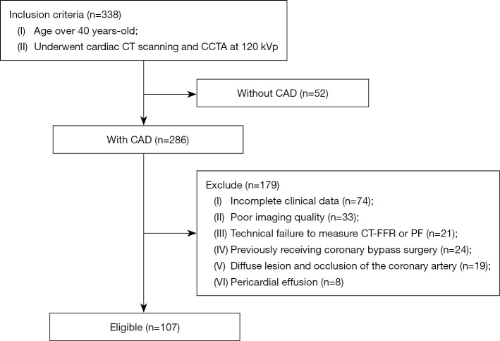
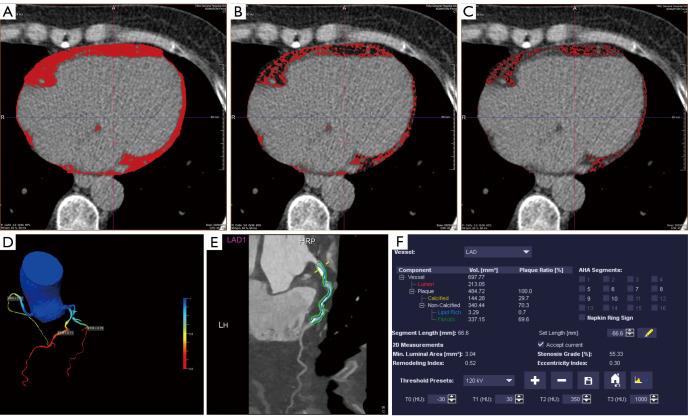
Methods: The clinical risk factors and imaging images of 107 CAD patients were retrospectively analyzed. Quantification of coronary artery calcium scores (CACS), PF, WAT, BAT, and pericardial fat attenuation (PFatt) were performed on non-contrast CT images. CCTA was used to evaluate myocardial ischemia and the presence of HRP. Fractional flow reserve derived from CCTA (CT-FFR) was performed in three major coronary arteries, with a threshold of ≤0.80 considered indicative of the presence of lesion-specific ischemia. HRP was defined as the presence of at least two of the four HRP features including positive remodeling (PR), low attenuation, napkin-ring sign (NRS), and spotty calcification. Mediator analysis was performed using Hayes (2018) Model-4.
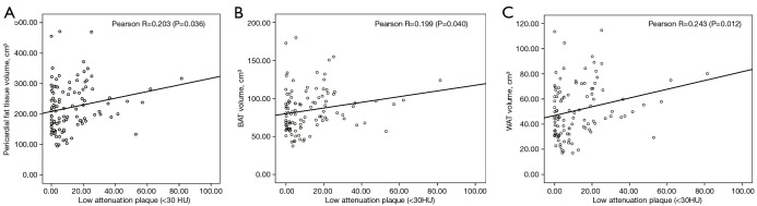
Results: A total of 107 CAD patients aged 65±8 years were included in this study. There were 49 patients (45.79%) with HRP and 57 patients (53.27%) with lesion-specific ischemia (CT-FFR ≤0.80). PF including WAT and BAT in the HRP group was significantly higher than that in the non-HRP group (PF: 241.28 vs. 204.94 cm3, P=0.005, WAT: 91.78 vs. 78.40 cm3, P=0.002, BAT: 56.25 vs. 46.71 cm3, P=0.008). Adding WAT to meaningful clinical risk factors and CACS increases the area under the curve (AUC) for HRP prediction {AUC [95% confidence interval (95% CI)]: 0.789 (0.692-0.887) vs. 0.655 (0.535-0.775), P<0.05}. Interestingly, adding PF to clinical risk factors and CACS decreased the AUC for the prediction of lesion-specific ischemia with no significant difference (P=0.083) [AUC (95% CI): 0.705 (0.592-0.817) vs. 0.796 (0.696-0.897), P<0.05]. Additionally, the mediation effect of PF accounted for 95.19% of the total effect of clinical risk factors on HRP (P<0.05).
Conclusions: PF is associated with HRP, and clinical risk factors increase the risk of plaque rupture by promoting PF volume accumulation, especially WAT, which may be a potential predictor of HRP.

 求助内容:
求助内容: 应助结果提醒方式:


