Xiao Hu, Jie-Jie Ding, Nian-Xia Qian, Xiao-Dong Liu
{"title":"克罗恩病肠系膜脂肪的计算机断层成像特征与炎症活动的相关性","authors":"Xiao Hu, Jie-Jie Ding, Nian-Xia Qian, Xiao-Dong Liu","doi":"10.21037/qims-2024-2606","DOIUrl":null,"url":null,"abstract":"<p><strong>Background: </strong>Creeping fat (CF), a characteristic structure of Crohn's disease (CD), is closely associated with surgery and prognosis but lacks a unified imaging assessment standard, and endoscopic and serological indicators have limitations in evaluating extra-intestinal lesions. The study aimed to explore the correlation between computed tomography (CT) value distribution changes of mesenteric-surrounding fat in CD and disease activity.</p><p><strong>Methods: </strong>In this study, we retrospectively analyzed CT enterography (CTE) images from 47 pathologically confirmed CD patients and 25 randomly selected controls with suspected inflammatory bowel disease (IBD). Quantitative measurements were obtained for mesenteric adipose tissue density (mean of CT values) along key anatomical landmarks including the mesenteric root, superior mesenteric artery, inferior mesenteric artery, perilesional regions, intestinal stricture, and adjacent branching vascular spaces. Other evaluated parameters included bowel wall thickness, CT attenuation, enhancement patterns, and CF grading. Qualitative evaluations were made by comparing with endoscopic, serological, and histopathological results and simplified CD activity index (CDAI) scores. A patient with concurrent anal fistula underwent magnetic resonance imaging (MRI) examination to compare its detection efficiency of fistula with that of CT examination.</p><p><strong>Results: </strong>Significant inter-group differences were found in non-contrast mesenteric fat attenuation, mean ΔCT (difference in Hounsfield units between contrast-enhanced CT and non-contrast CT scans) enhancement, bowel wall thickening, mucosal enhancement, CF grading, intestinal stricture, and serological parameters (P<0.05). The moderate-to-severe activity group had the highest mesenteric fat density in the venous phase [mean ΔCT >20 Hounsfield units (HU), P<0.05], especially around lesions. There was a positive correlation between mesenteric fat CT values and images of diseased bowel segments. The mean value of the venous phase ΔCT of the lesion and the mean of the intestinal wall venous phase ΔCT value and intestinal wall stratification were positively correlated (>0.6, P<0.05). ROC analysis showed that the venous-phase ΔCT of perilesional adipose tissue had excellent diagnostic performance [area under the curve (AUC) =0.964] for moderate-to-severe activity CD, with 95.8% sensitivity and 87.5% specificity. The diagnostic efficacy of the venous phase in the vascular space around the lesion ranked second (AUC =0.943). MRI showed superior detection of the anal fistula to that of CT in one patient. Multivariate analysis confirmed it as an independent predictor for moderate-to-severe active CD (P<0.05).</p><p><strong>Conclusions: </strong>Changes in mesenteric adipose tissue CT values and CF classification can distinguish CD from other IBD, suggesting their utility as another noninvasive diagnostic method for predicting the inflammatory activity of CD and evaluating the scope of surgery.</p>","PeriodicalId":54267,"journal":{"name":"Quantitative Imaging in Medicine and Surgery","volume":"15 8","pages":"6910-6922"},"PeriodicalIF":2.3000,"publicationDate":"2025-08-01","publicationTypes":"Journal Article","fieldsOfStudy":null,"isOpenAccess":false,"openAccessPdf":"https://www.ncbi.nlm.nih.gov/pmc/articles/PMC12332658/pdf/","citationCount":"0","resultStr":"{\"title\":\"Correlation between computed tomography imaging features of mesenteric fat and inflammatory activity in Crohn's disease.\",\"authors\":\"Xiao Hu, Jie-Jie Ding, Nian-Xia Qian, Xiao-Dong Liu\",\"doi\":\"10.21037/qims-2024-2606\",\"DOIUrl\":null,\"url\":null,\"abstract\":\"<p><strong>Background: </strong>Creeping fat (CF), a characteristic structure of Crohn's disease (CD), is closely associated with surgery and prognosis but lacks a unified imaging assessment standard, and endoscopic and serological indicators have limitations in evaluating extra-intestinal lesions. The study aimed to explore the correlation between computed tomography (CT) value distribution changes of mesenteric-surrounding fat in CD and disease activity.</p><p><strong>Methods: </strong>In this study, we retrospectively analyzed CT enterography (CTE) images from 47 pathologically confirmed CD patients and 25 randomly selected controls with suspected inflammatory bowel disease (IBD). Quantitative measurements were obtained for mesenteric adipose tissue density (mean of CT values) along key anatomical landmarks including the mesenteric root, superior mesenteric artery, inferior mesenteric artery, perilesional regions, intestinal stricture, and adjacent branching vascular spaces. Other evaluated parameters included bowel wall thickness, CT attenuation, enhancement patterns, and CF grading. Qualitative evaluations were made by comparing with endoscopic, serological, and histopathological results and simplified CD activity index (CDAI) scores. A patient with concurrent anal fistula underwent magnetic resonance imaging (MRI) examination to compare its detection efficiency of fistula with that of CT examination.</p><p><strong>Results: </strong>Significant inter-group differences were found in non-contrast mesenteric fat attenuation, mean ΔCT (difference in Hounsfield units between contrast-enhanced CT and non-contrast CT scans) enhancement, bowel wall thickening, mucosal enhancement, CF grading, intestinal stricture, and serological parameters (P<0.05). The moderate-to-severe activity group had the highest mesenteric fat density in the venous phase [mean ΔCT >20 Hounsfield units (HU), P<0.05], especially around lesions. There was a positive correlation between mesenteric fat CT values and images of diseased bowel segments. The mean value of the venous phase ΔCT of the lesion and the mean of the intestinal wall venous phase ΔCT value and intestinal wall stratification were positively correlated (>0.6, P<0.05). ROC analysis showed that the venous-phase ΔCT of perilesional adipose tissue had excellent diagnostic performance [area under the curve (AUC) =0.964] for moderate-to-severe activity CD, with 95.8% sensitivity and 87.5% specificity. The diagnostic efficacy of the venous phase in the vascular space around the lesion ranked second (AUC =0.943). MRI showed superior detection of the anal fistula to that of CT in one patient. Multivariate analysis confirmed it as an independent predictor for moderate-to-severe active CD (P<0.05).</p><p><strong>Conclusions: </strong>Changes in mesenteric adipose tissue CT values and CF classification can distinguish CD from other IBD, suggesting their utility as another noninvasive diagnostic method for predicting the inflammatory activity of CD and evaluating the scope of surgery.</p>\",\"PeriodicalId\":54267,\"journal\":{\"name\":\"Quantitative Imaging in Medicine and Surgery\",\"volume\":\"15 8\",\"pages\":\"6910-6922\"},\"PeriodicalIF\":2.3000,\"publicationDate\":\"2025-08-01\",\"publicationTypes\":\"Journal Article\",\"fieldsOfStudy\":null,\"isOpenAccess\":false,\"openAccessPdf\":\"https://www.ncbi.nlm.nih.gov/pmc/articles/PMC12332658/pdf/\",\"citationCount\":\"0\",\"resultStr\":null,\"platform\":\"Semanticscholar\",\"paperid\":null,\"PeriodicalName\":\"Quantitative Imaging in Medicine and Surgery\",\"FirstCategoryId\":\"3\",\"ListUrlMain\":\"https://doi.org/10.21037/qims-2024-2606\",\"RegionNum\":2,\"RegionCategory\":\"医学\",\"ArticlePicture\":[],\"TitleCN\":null,\"AbstractTextCN\":null,\"PMCID\":null,\"EPubDate\":\"2025/7/29 0:00:00\",\"PubModel\":\"Epub\",\"JCR\":\"Q2\",\"JCRName\":\"RADIOLOGY, NUCLEAR MEDICINE & MEDICAL IMAGING\",\"Score\":null,\"Total\":0}","platform":"Semanticscholar","paperid":null,"PeriodicalName":"Quantitative Imaging in Medicine and Surgery","FirstCategoryId":"3","ListUrlMain":"https://doi.org/10.21037/qims-2024-2606","RegionNum":2,"RegionCategory":"医学","ArticlePicture":[],"TitleCN":null,"AbstractTextCN":null,"PMCID":null,"EPubDate":"2025/7/29 0:00:00","PubModel":"Epub","JCR":"Q2","JCRName":"RADIOLOGY, NUCLEAR MEDICINE & MEDICAL IMAGING","Score":null,"Total":0}
Correlation between computed tomography imaging features of mesenteric fat and inflammatory activity in Crohn's disease.
Background: Creeping fat (CF), a characteristic structure of Crohn's disease (CD), is closely associated with surgery and prognosis but lacks a unified imaging assessment standard, and endoscopic and serological indicators have limitations in evaluating extra-intestinal lesions. The study aimed to explore the correlation between computed tomography (CT) value distribution changes of mesenteric-surrounding fat in CD and disease activity.
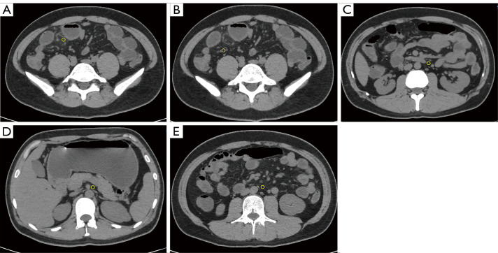
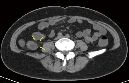
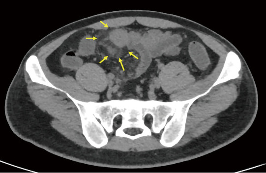
Methods: In this study, we retrospectively analyzed CT enterography (CTE) images from 47 pathologically confirmed CD patients and 25 randomly selected controls with suspected inflammatory bowel disease (IBD). Quantitative measurements were obtained for mesenteric adipose tissue density (mean of CT values) along key anatomical landmarks including the mesenteric root, superior mesenteric artery, inferior mesenteric artery, perilesional regions, intestinal stricture, and adjacent branching vascular spaces. Other evaluated parameters included bowel wall thickness, CT attenuation, enhancement patterns, and CF grading. Qualitative evaluations were made by comparing with endoscopic, serological, and histopathological results and simplified CD activity index (CDAI) scores. A patient with concurrent anal fistula underwent magnetic resonance imaging (MRI) examination to compare its detection efficiency of fistula with that of CT examination.
Results: Significant inter-group differences were found in non-contrast mesenteric fat attenuation, mean ΔCT (difference in Hounsfield units between contrast-enhanced CT and non-contrast CT scans) enhancement, bowel wall thickening, mucosal enhancement, CF grading, intestinal stricture, and serological parameters (P<0.05). The moderate-to-severe activity group had the highest mesenteric fat density in the venous phase [mean ΔCT >20 Hounsfield units (HU), P<0.05], especially around lesions. There was a positive correlation between mesenteric fat CT values and images of diseased bowel segments. The mean value of the venous phase ΔCT of the lesion and the mean of the intestinal wall venous phase ΔCT value and intestinal wall stratification were positively correlated (>0.6, P<0.05). ROC analysis showed that the venous-phase ΔCT of perilesional adipose tissue had excellent diagnostic performance [area under the curve (AUC) =0.964] for moderate-to-severe activity CD, with 95.8% sensitivity and 87.5% specificity. The diagnostic efficacy of the venous phase in the vascular space around the lesion ranked second (AUC =0.943). MRI showed superior detection of the anal fistula to that of CT in one patient. Multivariate analysis confirmed it as an independent predictor for moderate-to-severe active CD (P<0.05).
Conclusions: Changes in mesenteric adipose tissue CT values and CF classification can distinguish CD from other IBD, suggesting their utility as another noninvasive diagnostic method for predicting the inflammatory activity of CD and evaluating the scope of surgery.

 求助内容:
求助内容: 应助结果提醒方式:


