{"title":"细胞角蛋白19阳性肝细胞癌与常规肝细胞癌及肝内胆管癌的ct影像特征比较。","authors":"Hongbin Zhang, Lintao Chen, Jing Zhang, Ziqian Li, Yifan Wang, Fangyu Sun, Wenting Du, Xiuming Zhang, Wenjie Liang","doi":"10.21037/qims-24-914","DOIUrl":null,"url":null,"abstract":"<p><strong>Background: </strong>Cytokeratin 19-positive hepatocellular carcinoma (CK19<sup>+</sup> HCC) is an uncommon subtype of hepatocellular carcinoma (HCC). The purpose of this study was to identify radiological characteristics with diagnostic value for CK19<sup>+</sup> HCC.</p><p><strong>Methods: </strong>This was a case-control study. A retrospective analysis of 104 patients with surgically resected, pathologically confirmed CK19<sup>+</sup> HCC was conducted. The contrast-enhanced computed tomography characteristics of the enrolled patients were assessed, and differences in characteristics between groups were identified by statistical analysis. A multivariate logistic regression model was established to identify CK19<sup>+</sup> HCC, and receiver operating characteristic curves were plotted to evaluate the diagnostic performance of the model.</p><p><strong>Results: </strong>The univariate analysis revealed that the frequency of regular morphology (55.8% <i>vs.</i> 35.6%, P<0.001), hypodensity (99.0% <i>vs.</i> 91.8%, P=0.010), intratumoral necrosis (61.5% <i>vs.</i> 25.0%, P<0.001), heterogeneous enhancement (96.2% <i>vs.</i> 86.5%, P=0.008), peripheral washout (5.8% <i>vs.</i> 1.4%, P=0.031), non-peripheral washout (88.5% <i>vs.</i> 45.7%, P<0.001), Liver Imaging Reporting and Data System category 5 (67.3% <i>vs.</i> 40.4%, P<0.001), and Liver Imaging Reporting and Data System - Category tumor in vein (LR-TIV) (16.3% <i>vs.</i> 2.4%, P<0.001) were significantly higher in CK19<sup>+</sup> HCC than the non-CK19+ hepatic tumor patients. Conversely, the incidence of rim enhancement in the arterial phase (7.7% <i>vs.</i> 22.6%, P=0.001), transient hepatic attenuation difference (THAD; 4.8% <i>vs.</i> 23.1%, P<0.001), pseudocapsule formation (12.5% <i>vs.</i> 23.6%, P=0.021), progressive enhancement (5.8% <i>vs.</i> 50.5%, P<0.001), and lymphadenopathy (9.6% <i>vs.</i> 24.5%, P=0.002) was significantly lower in the CK19<sup>+</sup> HCC than the non-CK19<sup>+</sup> hepatic tumor patients. The multivariate analysis identified intratumoral necrosis, THAD, pseudocapsule formation, progressive enhancement, and LR-TIV as independent predictors of CK19+ HCC (P<0.05). The joint prediction model had an area under the curve of 0.867 in terms of its ability to detect CK19<sup>+</sup> HCC, and a sensitivity of 88.46% and a specificity of 69.71%.</p><p><strong>Conclusions: </strong>CK19<sup>+</sup> HCC is characterized by an increased prevalence of intratumoral necrosis and LR-TIV, as well as a lower incidence of THAD, pseudocapsule formation, and progressive enhancement, which collectively contribute to the identification of this HCC variant.</p>","PeriodicalId":54267,"journal":{"name":"Quantitative Imaging in Medicine and Surgery","volume":"15 8","pages":"7470-7482"},"PeriodicalIF":2.3000,"publicationDate":"2025-08-01","publicationTypes":"Journal Article","fieldsOfStudy":null,"isOpenAccess":false,"openAccessPdf":"https://www.ncbi.nlm.nih.gov/pmc/articles/PMC12332676/pdf/","citationCount":"0","resultStr":"{\"title\":\"Comparing the computed tomography radiologic features of cytokeratin 19-positive hepatocellular carcinoma to those of conventional hepatocellular carcinoma and intrahepatic cholangiocarcinoma.\",\"authors\":\"Hongbin Zhang, Lintao Chen, Jing Zhang, Ziqian Li, Yifan Wang, Fangyu Sun, Wenting Du, Xiuming Zhang, Wenjie Liang\",\"doi\":\"10.21037/qims-24-914\",\"DOIUrl\":null,\"url\":null,\"abstract\":\"<p><strong>Background: </strong>Cytokeratin 19-positive hepatocellular carcinoma (CK19<sup>+</sup> HCC) is an uncommon subtype of hepatocellular carcinoma (HCC). The purpose of this study was to identify radiological characteristics with diagnostic value for CK19<sup>+</sup> HCC.</p><p><strong>Methods: </strong>This was a case-control study. A retrospective analysis of 104 patients with surgically resected, pathologically confirmed CK19<sup>+</sup> HCC was conducted. The contrast-enhanced computed tomography characteristics of the enrolled patients were assessed, and differences in characteristics between groups were identified by statistical analysis. A multivariate logistic regression model was established to identify CK19<sup>+</sup> HCC, and receiver operating characteristic curves were plotted to evaluate the diagnostic performance of the model.</p><p><strong>Results: </strong>The univariate analysis revealed that the frequency of regular morphology (55.8% <i>vs.</i> 35.6%, P<0.001), hypodensity (99.0% <i>vs.</i> 91.8%, P=0.010), intratumoral necrosis (61.5% <i>vs.</i> 25.0%, P<0.001), heterogeneous enhancement (96.2% <i>vs.</i> 86.5%, P=0.008), peripheral washout (5.8% <i>vs.</i> 1.4%, P=0.031), non-peripheral washout (88.5% <i>vs.</i> 45.7%, P<0.001), Liver Imaging Reporting and Data System category 5 (67.3% <i>vs.</i> 40.4%, P<0.001), and Liver Imaging Reporting and Data System - Category tumor in vein (LR-TIV) (16.3% <i>vs.</i> 2.4%, P<0.001) were significantly higher in CK19<sup>+</sup> HCC than the non-CK19+ hepatic tumor patients. Conversely, the incidence of rim enhancement in the arterial phase (7.7% <i>vs.</i> 22.6%, P=0.001), transient hepatic attenuation difference (THAD; 4.8% <i>vs.</i> 23.1%, P<0.001), pseudocapsule formation (12.5% <i>vs.</i> 23.6%, P=0.021), progressive enhancement (5.8% <i>vs.</i> 50.5%, P<0.001), and lymphadenopathy (9.6% <i>vs.</i> 24.5%, P=0.002) was significantly lower in the CK19<sup>+</sup> HCC than the non-CK19<sup>+</sup> hepatic tumor patients. The multivariate analysis identified intratumoral necrosis, THAD, pseudocapsule formation, progressive enhancement, and LR-TIV as independent predictors of CK19+ HCC (P<0.05). The joint prediction model had an area under the curve of 0.867 in terms of its ability to detect CK19<sup>+</sup> HCC, and a sensitivity of 88.46% and a specificity of 69.71%.</p><p><strong>Conclusions: </strong>CK19<sup>+</sup> HCC is characterized by an increased prevalence of intratumoral necrosis and LR-TIV, as well as a lower incidence of THAD, pseudocapsule formation, and progressive enhancement, which collectively contribute to the identification of this HCC variant.</p>\",\"PeriodicalId\":54267,\"journal\":{\"name\":\"Quantitative Imaging in Medicine and Surgery\",\"volume\":\"15 8\",\"pages\":\"7470-7482\"},\"PeriodicalIF\":2.3000,\"publicationDate\":\"2025-08-01\",\"publicationTypes\":\"Journal Article\",\"fieldsOfStudy\":null,\"isOpenAccess\":false,\"openAccessPdf\":\"https://www.ncbi.nlm.nih.gov/pmc/articles/PMC12332676/pdf/\",\"citationCount\":\"0\",\"resultStr\":null,\"platform\":\"Semanticscholar\",\"paperid\":null,\"PeriodicalName\":\"Quantitative Imaging in Medicine and Surgery\",\"FirstCategoryId\":\"3\",\"ListUrlMain\":\"https://doi.org/10.21037/qims-24-914\",\"RegionNum\":2,\"RegionCategory\":\"医学\",\"ArticlePicture\":[],\"TitleCN\":null,\"AbstractTextCN\":null,\"PMCID\":null,\"EPubDate\":\"2025/7/29 0:00:00\",\"PubModel\":\"Epub\",\"JCR\":\"Q2\",\"JCRName\":\"RADIOLOGY, NUCLEAR MEDICINE & MEDICAL IMAGING\",\"Score\":null,\"Total\":0}","platform":"Semanticscholar","paperid":null,"PeriodicalName":"Quantitative Imaging in Medicine and Surgery","FirstCategoryId":"3","ListUrlMain":"https://doi.org/10.21037/qims-24-914","RegionNum":2,"RegionCategory":"医学","ArticlePicture":[],"TitleCN":null,"AbstractTextCN":null,"PMCID":null,"EPubDate":"2025/7/29 0:00:00","PubModel":"Epub","JCR":"Q2","JCRName":"RADIOLOGY, NUCLEAR MEDICINE & MEDICAL IMAGING","Score":null,"Total":0}
引用次数: 0
摘要
背景:细胞角蛋白19阳性肝细胞癌(CK19+ HCC)是一种罕见的肝细胞癌(HCC)亚型。本研究的目的是确定CK19+ HCC的放射学特征和诊断价值。方法:采用病例-对照研究。回顾性分析104例手术切除、病理证实的CK19+ HCC患者。评估入组患者的ct增强特征,并通过统计分析确定组间特征的差异。建立多元logistic回归模型识别CK19+ HCC,绘制受试者工作特征曲线评价模型的诊断效能。结果:单因素分析显示,P+肝癌患者的形态规则发生率(55.8%比35.6%,Pvs. 91.8%, P=0.010)、瘤内坏死发生率(61.5%比25.0%,Pvs. 86.5%, P=0.008)、外周洗脱(5.8%比1.4%,P=0.031)、非外周洗脱(88.5%比45.7%,Pvs. 40.4%, Pvs. 2.4%)高于非ck19 +肝肿瘤患者。相反,动脉期边缘增强的发生率(7.7% vs. 22.6%, P=0.001)、一过性肝衰减差(THAD;CK19+ HCC患者的进行性增强(5.8% vs. 50.5%, pv vs. 24.5%, P=0.002)显著低于非CK19+肝肿瘤患者。多变量分析发现,肿瘤内坏死、THAD、假包膜形成、进行性强化和LR-TIV是CK19+ HCC (P+ HCC)的独立预测因子,敏感性为88.46%,特异性为69.71%。结论:CK19+ HCC的特点是肿瘤内坏死和LR-TIV的发生率增加,THAD、假包膜形成和进行性强化的发生率较低,这些共同有助于识别这种HCC变体。
Comparing the computed tomography radiologic features of cytokeratin 19-positive hepatocellular carcinoma to those of conventional hepatocellular carcinoma and intrahepatic cholangiocarcinoma.
Background: Cytokeratin 19-positive hepatocellular carcinoma (CK19+ HCC) is an uncommon subtype of hepatocellular carcinoma (HCC). The purpose of this study was to identify radiological characteristics with diagnostic value for CK19+ HCC.
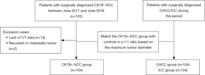
Methods: This was a case-control study. A retrospective analysis of 104 patients with surgically resected, pathologically confirmed CK19+ HCC was conducted. The contrast-enhanced computed tomography characteristics of the enrolled patients were assessed, and differences in characteristics between groups were identified by statistical analysis. A multivariate logistic regression model was established to identify CK19+ HCC, and receiver operating characteristic curves were plotted to evaluate the diagnostic performance of the model.
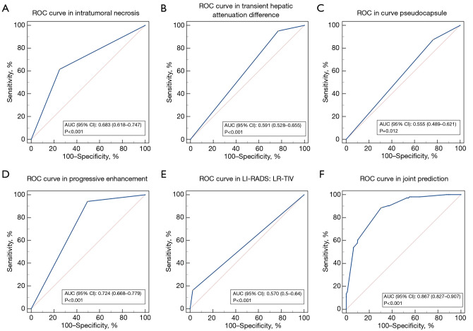
Results: The univariate analysis revealed that the frequency of regular morphology (55.8% vs. 35.6%, P<0.001), hypodensity (99.0% vs. 91.8%, P=0.010), intratumoral necrosis (61.5% vs. 25.0%, P<0.001), heterogeneous enhancement (96.2% vs. 86.5%, P=0.008), peripheral washout (5.8% vs. 1.4%, P=0.031), non-peripheral washout (88.5% vs. 45.7%, P<0.001), Liver Imaging Reporting and Data System category 5 (67.3% vs. 40.4%, P<0.001), and Liver Imaging Reporting and Data System - Category tumor in vein (LR-TIV) (16.3% vs. 2.4%, P<0.001) were significantly higher in CK19+ HCC than the non-CK19+ hepatic tumor patients. Conversely, the incidence of rim enhancement in the arterial phase (7.7% vs. 22.6%, P=0.001), transient hepatic attenuation difference (THAD; 4.8% vs. 23.1%, P<0.001), pseudocapsule formation (12.5% vs. 23.6%, P=0.021), progressive enhancement (5.8% vs. 50.5%, P<0.001), and lymphadenopathy (9.6% vs. 24.5%, P=0.002) was significantly lower in the CK19+ HCC than the non-CK19+ hepatic tumor patients. The multivariate analysis identified intratumoral necrosis, THAD, pseudocapsule formation, progressive enhancement, and LR-TIV as independent predictors of CK19+ HCC (P<0.05). The joint prediction model had an area under the curve of 0.867 in terms of its ability to detect CK19+ HCC, and a sensitivity of 88.46% and a specificity of 69.71%.
Conclusions: CK19+ HCC is characterized by an increased prevalence of intratumoral necrosis and LR-TIV, as well as a lower incidence of THAD, pseudocapsule formation, and progressive enhancement, which collectively contribute to the identification of this HCC variant.

 求助内容:
求助内容: 应助结果提醒方式:


