Seher Koksaldi, Mustafa Kayabasi, Omer Karti, Ali Osman Saatci
{"title":"黄斑的临床解剖。","authors":"Seher Koksaldi, Mustafa Kayabasi, Omer Karti, Ali Osman Saatci","doi":"10.51329/mehdiophthal1520","DOIUrl":null,"url":null,"abstract":"<p><strong>Background: </strong>The macula is located at the center of the retina and is crucial for high-resolution color vision. Its complex anatomical structure supports a dense array of cone photoreceptors and specialized neuronal pathways essential for central vision. A thorough understanding of macular microanatomy is vital for accurate interpretation of retinal imaging and effective management of macular diseases. This narrative review provides a detailed and integrative overview of macular anatomy, emphasizing clinically relevant microanatomical features and their implications in retinal imaging and macular disease management.</p><p><strong>Methods: </strong>A PubMed/MEDLINE search was performed using relevant keywords (e.g., \"anatomy,\" \"fovea,\" \"foveal avascular zone,\" \"foveola,\" \"Henle fiber layer,\" \"macula,\" \"macular anatomy,\" \"macula lutea,\" \"optical coherence tomography,\" \"parafovea,\" \"perifovea,\" and \"retina\") to identify English-language articles published up to February 28, 2025. The reference lists of the included papers were manually reviewed to identify additional relevant sources. The review considered a wide range of study types, including clinical trials, systematic and narrative reviews, meta-analyses, observational studies, case series, and experimental animal studies.</p><p><strong>Results: </strong>This review highlights the remarkable characteristics of the fovea and foveola, which are densely packed with cone photoreceptors, making them uniquely suited for sharp vision. The surrounding parafoveal and perifoveal regions offer critical structural and functional support, while the Henle fiber layer facilitates the oblique course of photoreceptor axons, further refining central vision. Moreover, high-resolution optical coherence tomography has revolutionized visualization of the macular architecture, enabling a detailed assessment of previously undetectable retinal layers. This review explores key anatomical features, such as the foveal avascular zone, precise photoreceptor organization, and the role of Muller glial cells, in the context of high-resolution imaging. These associations between anatomy and imaging enhance diagnostic precision and may inform targeted treatment approaches for macular diseases.</p><p><strong>Conclusions: </strong>Comprehensive knowledge of macular anatomy is crucial for the accurate interpretation of retinal imaging and management of central retinal disorders. The bridging of classic histological findings with modern imaging enhances comprehension of the healthy macula and the detection and management of pathological changes. This review serves as a practical anatomical reference for clinicians and researchers in macular diagnostics and therapeutics. Further studies are warranted to explore how emerging imaging technologies can enhance early detection and treatment strategies for macular disorders.</p>","PeriodicalId":36524,"journal":{"name":"Medical Hypothesis, Discovery, and Innovation in Ophthalmology","volume":"14 2","pages":"17-27"},"PeriodicalIF":0.0000,"publicationDate":"2025-07-31","publicationTypes":"Journal Article","fieldsOfStudy":null,"isOpenAccess":false,"openAccessPdf":"https://www.ncbi.nlm.nih.gov/pmc/articles/PMC12330045/pdf/","citationCount":"0","resultStr":"{\"title\":\"Clinical anatomy of the macula.\",\"authors\":\"Seher Koksaldi, Mustafa Kayabasi, Omer Karti, Ali Osman Saatci\",\"doi\":\"10.51329/mehdiophthal1520\",\"DOIUrl\":null,\"url\":null,\"abstract\":\"<p><strong>Background: </strong>The macula is located at the center of the retina and is crucial for high-resolution color vision. Its complex anatomical structure supports a dense array of cone photoreceptors and specialized neuronal pathways essential for central vision. A thorough understanding of macular microanatomy is vital for accurate interpretation of retinal imaging and effective management of macular diseases. This narrative review provides a detailed and integrative overview of macular anatomy, emphasizing clinically relevant microanatomical features and their implications in retinal imaging and macular disease management.</p><p><strong>Methods: </strong>A PubMed/MEDLINE search was performed using relevant keywords (e.g., \\\"anatomy,\\\" \\\"fovea,\\\" \\\"foveal avascular zone,\\\" \\\"foveola,\\\" \\\"Henle fiber layer,\\\" \\\"macula,\\\" \\\"macular anatomy,\\\" \\\"macula lutea,\\\" \\\"optical coherence tomography,\\\" \\\"parafovea,\\\" \\\"perifovea,\\\" and \\\"retina\\\") to identify English-language articles published up to February 28, 2025. The reference lists of the included papers were manually reviewed to identify additional relevant sources. The review considered a wide range of study types, including clinical trials, systematic and narrative reviews, meta-analyses, observational studies, case series, and experimental animal studies.</p><p><strong>Results: </strong>This review highlights the remarkable characteristics of the fovea and foveola, which are densely packed with cone photoreceptors, making them uniquely suited for sharp vision. The surrounding parafoveal and perifoveal regions offer critical structural and functional support, while the Henle fiber layer facilitates the oblique course of photoreceptor axons, further refining central vision. Moreover, high-resolution optical coherence tomography has revolutionized visualization of the macular architecture, enabling a detailed assessment of previously undetectable retinal layers. This review explores key anatomical features, such as the foveal avascular zone, precise photoreceptor organization, and the role of Muller glial cells, in the context of high-resolution imaging. These associations between anatomy and imaging enhance diagnostic precision and may inform targeted treatment approaches for macular diseases.</p><p><strong>Conclusions: </strong>Comprehensive knowledge of macular anatomy is crucial for the accurate interpretation of retinal imaging and management of central retinal disorders. The bridging of classic histological findings with modern imaging enhances comprehension of the healthy macula and the detection and management of pathological changes. This review serves as a practical anatomical reference for clinicians and researchers in macular diagnostics and therapeutics. Further studies are warranted to explore how emerging imaging technologies can enhance early detection and treatment strategies for macular disorders.</p>\",\"PeriodicalId\":36524,\"journal\":{\"name\":\"Medical Hypothesis, Discovery, and Innovation in Ophthalmology\",\"volume\":\"14 2\",\"pages\":\"17-27\"},\"PeriodicalIF\":0.0000,\"publicationDate\":\"2025-07-31\",\"publicationTypes\":\"Journal Article\",\"fieldsOfStudy\":null,\"isOpenAccess\":false,\"openAccessPdf\":\"https://www.ncbi.nlm.nih.gov/pmc/articles/PMC12330045/pdf/\",\"citationCount\":\"0\",\"resultStr\":null,\"platform\":\"Semanticscholar\",\"paperid\":null,\"PeriodicalName\":\"Medical Hypothesis, Discovery, and Innovation in Ophthalmology\",\"FirstCategoryId\":\"1085\",\"ListUrlMain\":\"https://doi.org/10.51329/mehdiophthal1520\",\"RegionNum\":0,\"RegionCategory\":null,\"ArticlePicture\":[],\"TitleCN\":null,\"AbstractTextCN\":null,\"PMCID\":null,\"EPubDate\":\"2025/1/1 0:00:00\",\"PubModel\":\"eCollection\",\"JCR\":\"Q2\",\"JCRName\":\"Medicine\",\"Score\":null,\"Total\":0}","platform":"Semanticscholar","paperid":null,"PeriodicalName":"Medical Hypothesis, Discovery, and Innovation in Ophthalmology","FirstCategoryId":"1085","ListUrlMain":"https://doi.org/10.51329/mehdiophthal1520","RegionNum":0,"RegionCategory":null,"ArticlePicture":[],"TitleCN":null,"AbstractTextCN":null,"PMCID":null,"EPubDate":"2025/1/1 0:00:00","PubModel":"eCollection","JCR":"Q2","JCRName":"Medicine","Score":null,"Total":0}
Background: The macula is located at the center of the retina and is crucial for high-resolution color vision. Its complex anatomical structure supports a dense array of cone photoreceptors and specialized neuronal pathways essential for central vision. A thorough understanding of macular microanatomy is vital for accurate interpretation of retinal imaging and effective management of macular diseases. This narrative review provides a detailed and integrative overview of macular anatomy, emphasizing clinically relevant microanatomical features and their implications in retinal imaging and macular disease management.
Methods: A PubMed/MEDLINE search was performed using relevant keywords (e.g., "anatomy," "fovea," "foveal avascular zone," "foveola," "Henle fiber layer," "macula," "macular anatomy," "macula lutea," "optical coherence tomography," "parafovea," "perifovea," and "retina") to identify English-language articles published up to February 28, 2025. The reference lists of the included papers were manually reviewed to identify additional relevant sources. The review considered a wide range of study types, including clinical trials, systematic and narrative reviews, meta-analyses, observational studies, case series, and experimental animal studies.
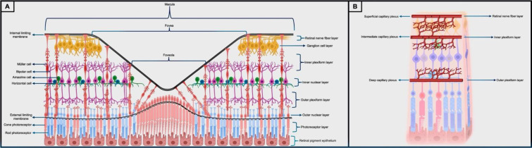
Results: This review highlights the remarkable characteristics of the fovea and foveola, which are densely packed with cone photoreceptors, making them uniquely suited for sharp vision. The surrounding parafoveal and perifoveal regions offer critical structural and functional support, while the Henle fiber layer facilitates the oblique course of photoreceptor axons, further refining central vision. Moreover, high-resolution optical coherence tomography has revolutionized visualization of the macular architecture, enabling a detailed assessment of previously undetectable retinal layers. This review explores key anatomical features, such as the foveal avascular zone, precise photoreceptor organization, and the role of Muller glial cells, in the context of high-resolution imaging. These associations between anatomy and imaging enhance diagnostic precision and may inform targeted treatment approaches for macular diseases.
Conclusions: Comprehensive knowledge of macular anatomy is crucial for the accurate interpretation of retinal imaging and management of central retinal disorders. The bridging of classic histological findings with modern imaging enhances comprehension of the healthy macula and the detection and management of pathological changes. This review serves as a practical anatomical reference for clinicians and researchers in macular diagnostics and therapeutics. Further studies are warranted to explore how emerging imaging technologies can enhance early detection and treatment strategies for macular disorders.

 求助内容:
求助内容: 应助结果提醒方式:


