{"title":"三维计算机断层成像定量评估成人脊柱畸形手术中腹腔和肠系膜上动脉直径。","authors":"Yasushi Iijima, Toshiaki Kotani, Tsuyoshi Sakuma, Tsutomu Akazawa, Shunji Kishida, Keisuke Ueno, Shohei Ise, Shuhei Ohyama, Shuhei Iwata, Masaya Mizutani, Kotaro Sakashita, Takahiro Sunami, Shun Okuwaki, Yosuke Ogata, Yasuhiro Shiga, Shohei Minami, Seiji Ohtori","doi":"10.22603/ssrr.2024-0228","DOIUrl":null,"url":null,"abstract":"<p><strong>Introduction: </strong>Acute celiac artery compression syndrome occurs after corrective surgery for adult spinal deformity. It occurs due to ischemic abdominal organ necrosis, caused by compression of the celiac artery (CA) and superior mesenteric artery by the median arcuate ligament. There are no studies measuring the extent of CA or superior mesenteric artery stenosis. Therefore, this study aimed to investigate stenotic changes in the CA after adult spinal deformity surgery.</p><p><strong>Methods: </strong>We obtained contrast-enhanced computed tomography scans for 21 pre-and postoperative patients with adult spinal deformity. Three-dimensional reconstruction computed tomography measured the degree of stenosis in the CA trunks. Stenosis was considered worse if it progressed from being less than 35% before surgery to over 50% afterward. This study investigated the relationship between worsening CA stenosis and the median arcuate ligament crossing the proximal portion of the celiac axis (median arcuate ligament overlap) or the distance between the median arcuate ligament and the anterior edge of the vertebra (DMV). Change in spinal parameters was defined as differences between pre- and postoperative values.</p><p><strong>Results: </strong>The average stenosis degree in the CA was 9.4%±11.4% pre-operatively, which increased to 25.1%±21.8% post-operatively (<i>P</i>=0.002). In contrast, the stenosis degree in the superior mesenteric artery was 5.6%±7.1% before and 7.9%±10.2% after surgery (<i>P</i>=0.177). CA stenosis worsened in four patients (19.0%), which was significantly associated with preoperative median arcuate ligament overlap (<i>P</i>=0.012) and ΔDMV (<i>P</i><0.001).</p><p><strong>Conclusions: </strong>Nineteen percent of patients undergoing adult spinal deformity correction surgery experienced worsened CA stenosis. Risk factors were preoperative median arcuate ligament overlap and DMV shortening during adult spinal deformity correction surgery. Moreover, patients with preoperative CA stenosis and median arcuate ligament overlap were at risk for acute celiac artery compression syndrome following adult spinal deformity surgery.</p>","PeriodicalId":22253,"journal":{"name":"Spine Surgery and Related Research","volume":"9 4","pages":"477-484"},"PeriodicalIF":1.2000,"publicationDate":"2024-12-20","publicationTypes":"Journal Article","fieldsOfStudy":null,"isOpenAccess":false,"openAccessPdf":"https://www.ncbi.nlm.nih.gov/pmc/articles/PMC12330376/pdf/","citationCount":"0","resultStr":"{\"title\":\"Quantitative Assessment of Celiac and Superior Mesenteric Artery Diameters in Adult Spinal Deformity Surgery Using Three-dimensional Computed Tomography.\",\"authors\":\"Yasushi Iijima, Toshiaki Kotani, Tsuyoshi Sakuma, Tsutomu Akazawa, Shunji Kishida, Keisuke Ueno, Shohei Ise, Shuhei Ohyama, Shuhei Iwata, Masaya Mizutani, Kotaro Sakashita, Takahiro Sunami, Shun Okuwaki, Yosuke Ogata, Yasuhiro Shiga, Shohei Minami, Seiji Ohtori\",\"doi\":\"10.22603/ssrr.2024-0228\",\"DOIUrl\":null,\"url\":null,\"abstract\":\"<p><strong>Introduction: </strong>Acute celiac artery compression syndrome occurs after corrective surgery for adult spinal deformity. It occurs due to ischemic abdominal organ necrosis, caused by compression of the celiac artery (CA) and superior mesenteric artery by the median arcuate ligament. There are no studies measuring the extent of CA or superior mesenteric artery stenosis. Therefore, this study aimed to investigate stenotic changes in the CA after adult spinal deformity surgery.</p><p><strong>Methods: </strong>We obtained contrast-enhanced computed tomography scans for 21 pre-and postoperative patients with adult spinal deformity. Three-dimensional reconstruction computed tomography measured the degree of stenosis in the CA trunks. Stenosis was considered worse if it progressed from being less than 35% before surgery to over 50% afterward. This study investigated the relationship between worsening CA stenosis and the median arcuate ligament crossing the proximal portion of the celiac axis (median arcuate ligament overlap) or the distance between the median arcuate ligament and the anterior edge of the vertebra (DMV). Change in spinal parameters was defined as differences between pre- and postoperative values.</p><p><strong>Results: </strong>The average stenosis degree in the CA was 9.4%±11.4% pre-operatively, which increased to 25.1%±21.8% post-operatively (<i>P</i>=0.002). In contrast, the stenosis degree in the superior mesenteric artery was 5.6%±7.1% before and 7.9%±10.2% after surgery (<i>P</i>=0.177). CA stenosis worsened in four patients (19.0%), which was significantly associated with preoperative median arcuate ligament overlap (<i>P</i>=0.012) and ΔDMV (<i>P</i><0.001).</p><p><strong>Conclusions: </strong>Nineteen percent of patients undergoing adult spinal deformity correction surgery experienced worsened CA stenosis. Risk factors were preoperative median arcuate ligament overlap and DMV shortening during adult spinal deformity correction surgery. Moreover, patients with preoperative CA stenosis and median arcuate ligament overlap were at risk for acute celiac artery compression syndrome following adult spinal deformity surgery.</p>\",\"PeriodicalId\":22253,\"journal\":{\"name\":\"Spine Surgery and Related Research\",\"volume\":\"9 4\",\"pages\":\"477-484\"},\"PeriodicalIF\":1.2000,\"publicationDate\":\"2024-12-20\",\"publicationTypes\":\"Journal Article\",\"fieldsOfStudy\":null,\"isOpenAccess\":false,\"openAccessPdf\":\"https://www.ncbi.nlm.nih.gov/pmc/articles/PMC12330376/pdf/\",\"citationCount\":\"0\",\"resultStr\":null,\"platform\":\"Semanticscholar\",\"paperid\":null,\"PeriodicalName\":\"Spine Surgery and Related Research\",\"FirstCategoryId\":\"1085\",\"ListUrlMain\":\"https://doi.org/10.22603/ssrr.2024-0228\",\"RegionNum\":0,\"RegionCategory\":null,\"ArticlePicture\":[],\"TitleCN\":null,\"AbstractTextCN\":null,\"PMCID\":null,\"EPubDate\":\"2025/7/27 0:00:00\",\"PubModel\":\"eCollection\",\"JCR\":\"Q3\",\"JCRName\":\"SURGERY\",\"Score\":null,\"Total\":0}","platform":"Semanticscholar","paperid":null,"PeriodicalName":"Spine Surgery and Related Research","FirstCategoryId":"1085","ListUrlMain":"https://doi.org/10.22603/ssrr.2024-0228","RegionNum":0,"RegionCategory":null,"ArticlePicture":[],"TitleCN":null,"AbstractTextCN":null,"PMCID":null,"EPubDate":"2025/7/27 0:00:00","PubModel":"eCollection","JCR":"Q3","JCRName":"SURGERY","Score":null,"Total":0}
Quantitative Assessment of Celiac and Superior Mesenteric Artery Diameters in Adult Spinal Deformity Surgery Using Three-dimensional Computed Tomography.
Introduction: Acute celiac artery compression syndrome occurs after corrective surgery for adult spinal deformity. It occurs due to ischemic abdominal organ necrosis, caused by compression of the celiac artery (CA) and superior mesenteric artery by the median arcuate ligament. There are no studies measuring the extent of CA or superior mesenteric artery stenosis. Therefore, this study aimed to investigate stenotic changes in the CA after adult spinal deformity surgery.
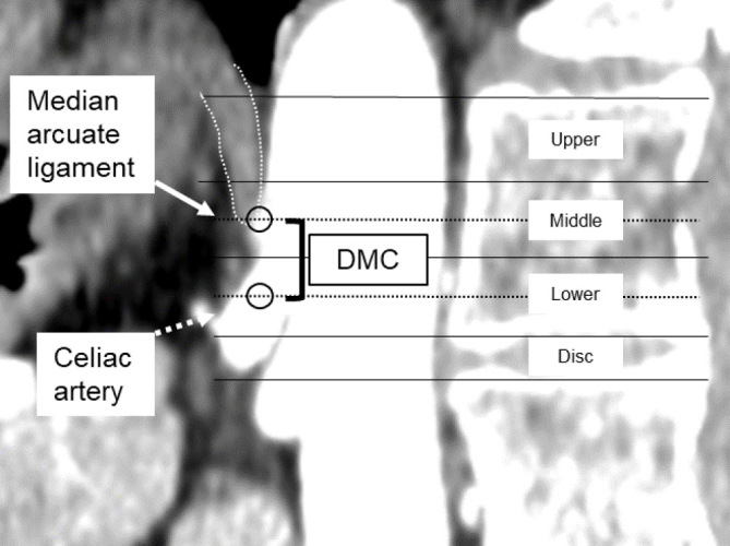
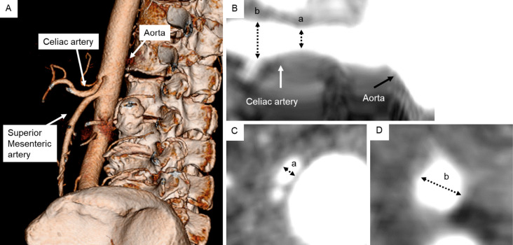
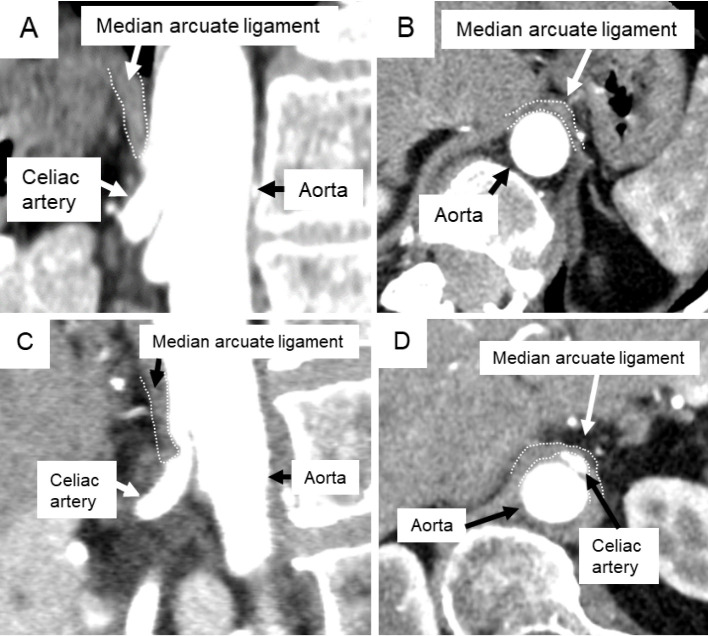
Methods: We obtained contrast-enhanced computed tomography scans for 21 pre-and postoperative patients with adult spinal deformity. Three-dimensional reconstruction computed tomography measured the degree of stenosis in the CA trunks. Stenosis was considered worse if it progressed from being less than 35% before surgery to over 50% afterward. This study investigated the relationship between worsening CA stenosis and the median arcuate ligament crossing the proximal portion of the celiac axis (median arcuate ligament overlap) or the distance between the median arcuate ligament and the anterior edge of the vertebra (DMV). Change in spinal parameters was defined as differences between pre- and postoperative values.
Results: The average stenosis degree in the CA was 9.4%±11.4% pre-operatively, which increased to 25.1%±21.8% post-operatively (P=0.002). In contrast, the stenosis degree in the superior mesenteric artery was 5.6%±7.1% before and 7.9%±10.2% after surgery (P=0.177). CA stenosis worsened in four patients (19.0%), which was significantly associated with preoperative median arcuate ligament overlap (P=0.012) and ΔDMV (P<0.001).
Conclusions: Nineteen percent of patients undergoing adult spinal deformity correction surgery experienced worsened CA stenosis. Risk factors were preoperative median arcuate ligament overlap and DMV shortening during adult spinal deformity correction surgery. Moreover, patients with preoperative CA stenosis and median arcuate ligament overlap were at risk for acute celiac artery compression syndrome following adult spinal deformity surgery.

 求助内容:
求助内容: 应助结果提醒方式:


