Mengmeng Dou, Jinfeng Yang, Jiaoran Li, Jianmin Li, Zhenzhong Li, Xianhao Shao, Xia Su, Jing Li, Guanghui Wang, Kun Cheng
{"title":"一种新的纳米技术用于关节附近骨内血管-管道复合体的成像及相关的临床试验研究。","authors":"Mengmeng Dou, Jinfeng Yang, Jiaoran Li, Jianmin Li, Zhenzhong Li, Xianhao Shao, Xia Su, Jing Li, Guanghui Wang, Kun Cheng","doi":"10.1111/os.70149","DOIUrl":null,"url":null,"abstract":"<p><strong>Objective: </strong>The objective of this study is to achieve distinct visualization of juxta-articular intraosseous microvessels, a novel nanoimaging methodology in which superparamagnetic iron oxide nanoparticles and meglumine diatrizoate (MD) are used cooperatively was implemented.</p><p><strong>Methods: </strong>A newly created composite of MD and Fe<sub>3</sub>O<sub>4</sub> nanoparticles (MD-Fe<sub>3</sub>O<sub>4</sub> NPs) was prepared as a contrast agent to achieve efficacious imaging of the juxta-articular intraosseous vasculature-canal complex (JIVCC). Scanning electron microscopy (SEM) and energy dispersive spectrum (EDS) were employed to observe the structural characteristics and binding stability of the MD-Fe<sub>3</sub>O<sub>4</sub> NPs. In 20 rabbits that received an injection of MD-Fe<sub>3</sub>O<sub>4</sub> NPs, 1-mm-thick computed tomography (CT) scanning was performed for radiographic assessment. Hematoxylin-eosin- and potassium ferrocyanide-stained sections from 10 sacrificed rabbits were used to observe the histological characteristics of JIVCC with MD-Fe<sub>3</sub>O<sub>4</sub> NPs, and the remaining 10 rabbits were utilized for a systemic safety evaluation. After a healthy volunteer received an MD-Fe<sub>3</sub>O<sub>4</sub> NP injection, we also performed CT scanning and related safety evaluations.</p><p><strong>Results: </strong>When the MD nanoparticles and amino-Fe<sub>3</sub>O<sub>4</sub> nanoparticles were mixed together, they aggregated into a stable compound structure according to microscopic observations and SEM-EDS verification. In 20 rabbits receiving MD-Fe<sub>3</sub>O<sub>4</sub> injections, 1-mm slice CT imaging demonstrated significantly enhanced visualization of the JIVCCs in magnet-placed knees compared to contralateral limbs (tibial JIVCC: p < 0.001; femoral JIVCC: p < 0.001), confirming MD-Fe<sub>3</sub>O<sub>4</sub> NPs as the efficacious magnetic contrast enhancer. The histological characteristics of MD-Fe<sub>3</sub>O<sub>4</sub> NPs in JIVCC were revealed. The levels of serum iron before and 4 and 72 h after MD-Fe<sub>3</sub>O<sub>4</sub> NP injection were 23.9 ± 2.13 μmol/L, 26.2 ± 2.30 μmol/L, and 24.9 ± 2.33 μmol/L, respectively, indicating that there was no significant difference in safety (p = 0.092). After a volunteer received MD-Fe<sub>3</sub>O<sub>4</sub> NPs via intravenous administration, the JIVCC was clearly visualized, laboratory tests of serum iron levels were normal, and no injection-related complications occurred.</p><p><strong>Conclusions: </strong>A novel compound nanoparticle, which achieved satisfactory overall outcomes, was implemented as an appropriate alternative for the discernible visualization of juxta-articular intraosseous microvessels. The nanotechnology utilized in this study may augment the clinical imaging methodology for the osseous vascular system.</p>","PeriodicalId":19566,"journal":{"name":"Orthopaedic Surgery","volume":" ","pages":"2744-2755"},"PeriodicalIF":2.1000,"publicationDate":"2025-09-01","publicationTypes":"Journal Article","fieldsOfStudy":null,"isOpenAccess":false,"openAccessPdf":"https://www.ncbi.nlm.nih.gov/pmc/articles/PMC12404870/pdf/","citationCount":"0","resultStr":"{\"title\":\"A New Nanotechnology for Imaging the Juxta-Articular Intraosseous Vasculature-Canal Complex and Related Clinical Pilot Studies.\",\"authors\":\"Mengmeng Dou, Jinfeng Yang, Jiaoran Li, Jianmin Li, Zhenzhong Li, Xianhao Shao, Xia Su, Jing Li, Guanghui Wang, Kun Cheng\",\"doi\":\"10.1111/os.70149\",\"DOIUrl\":null,\"url\":null,\"abstract\":\"<p><strong>Objective: </strong>The objective of this study is to achieve distinct visualization of juxta-articular intraosseous microvessels, a novel nanoimaging methodology in which superparamagnetic iron oxide nanoparticles and meglumine diatrizoate (MD) are used cooperatively was implemented.</p><p><strong>Methods: </strong>A newly created composite of MD and Fe<sub>3</sub>O<sub>4</sub> nanoparticles (MD-Fe<sub>3</sub>O<sub>4</sub> NPs) was prepared as a contrast agent to achieve efficacious imaging of the juxta-articular intraosseous vasculature-canal complex (JIVCC). Scanning electron microscopy (SEM) and energy dispersive spectrum (EDS) were employed to observe the structural characteristics and binding stability of the MD-Fe<sub>3</sub>O<sub>4</sub> NPs. In 20 rabbits that received an injection of MD-Fe<sub>3</sub>O<sub>4</sub> NPs, 1-mm-thick computed tomography (CT) scanning was performed for radiographic assessment. Hematoxylin-eosin- and potassium ferrocyanide-stained sections from 10 sacrificed rabbits were used to observe the histological characteristics of JIVCC with MD-Fe<sub>3</sub>O<sub>4</sub> NPs, and the remaining 10 rabbits were utilized for a systemic safety evaluation. After a healthy volunteer received an MD-Fe<sub>3</sub>O<sub>4</sub> NP injection, we also performed CT scanning and related safety evaluations.</p><p><strong>Results: </strong>When the MD nanoparticles and amino-Fe<sub>3</sub>O<sub>4</sub> nanoparticles were mixed together, they aggregated into a stable compound structure according to microscopic observations and SEM-EDS verification. In 20 rabbits receiving MD-Fe<sub>3</sub>O<sub>4</sub> injections, 1-mm slice CT imaging demonstrated significantly enhanced visualization of the JIVCCs in magnet-placed knees compared to contralateral limbs (tibial JIVCC: p < 0.001; femoral JIVCC: p < 0.001), confirming MD-Fe<sub>3</sub>O<sub>4</sub> NPs as the efficacious magnetic contrast enhancer. The histological characteristics of MD-Fe<sub>3</sub>O<sub>4</sub> NPs in JIVCC were revealed. The levels of serum iron before and 4 and 72 h after MD-Fe<sub>3</sub>O<sub>4</sub> NP injection were 23.9 ± 2.13 μmol/L, 26.2 ± 2.30 μmol/L, and 24.9 ± 2.33 μmol/L, respectively, indicating that there was no significant difference in safety (p = 0.092). After a volunteer received MD-Fe<sub>3</sub>O<sub>4</sub> NPs via intravenous administration, the JIVCC was clearly visualized, laboratory tests of serum iron levels were normal, and no injection-related complications occurred.</p><p><strong>Conclusions: </strong>A novel compound nanoparticle, which achieved satisfactory overall outcomes, was implemented as an appropriate alternative for the discernible visualization of juxta-articular intraosseous microvessels. The nanotechnology utilized in this study may augment the clinical imaging methodology for the osseous vascular system.</p>\",\"PeriodicalId\":19566,\"journal\":{\"name\":\"Orthopaedic Surgery\",\"volume\":\" \",\"pages\":\"2744-2755\"},\"PeriodicalIF\":2.1000,\"publicationDate\":\"2025-09-01\",\"publicationTypes\":\"Journal Article\",\"fieldsOfStudy\":null,\"isOpenAccess\":false,\"openAccessPdf\":\"https://www.ncbi.nlm.nih.gov/pmc/articles/PMC12404870/pdf/\",\"citationCount\":\"0\",\"resultStr\":null,\"platform\":\"Semanticscholar\",\"paperid\":null,\"PeriodicalName\":\"Orthopaedic Surgery\",\"FirstCategoryId\":\"3\",\"ListUrlMain\":\"https://doi.org/10.1111/os.70149\",\"RegionNum\":2,\"RegionCategory\":\"医学\",\"ArticlePicture\":[],\"TitleCN\":null,\"AbstractTextCN\":null,\"PMCID\":null,\"EPubDate\":\"2025/8/11 0:00:00\",\"PubModel\":\"Epub\",\"JCR\":\"Q2\",\"JCRName\":\"ORTHOPEDICS\",\"Score\":null,\"Total\":0}","platform":"Semanticscholar","paperid":null,"PeriodicalName":"Orthopaedic Surgery","FirstCategoryId":"3","ListUrlMain":"https://doi.org/10.1111/os.70149","RegionNum":2,"RegionCategory":"医学","ArticlePicture":[],"TitleCN":null,"AbstractTextCN":null,"PMCID":null,"EPubDate":"2025/8/11 0:00:00","PubModel":"Epub","JCR":"Q2","JCRName":"ORTHOPEDICS","Score":null,"Total":0}
A New Nanotechnology for Imaging the Juxta-Articular Intraosseous Vasculature-Canal Complex and Related Clinical Pilot Studies.
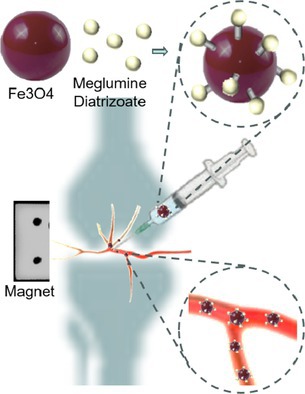
Objective: The objective of this study is to achieve distinct visualization of juxta-articular intraosseous microvessels, a novel nanoimaging methodology in which superparamagnetic iron oxide nanoparticles and meglumine diatrizoate (MD) are used cooperatively was implemented.
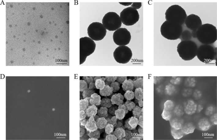
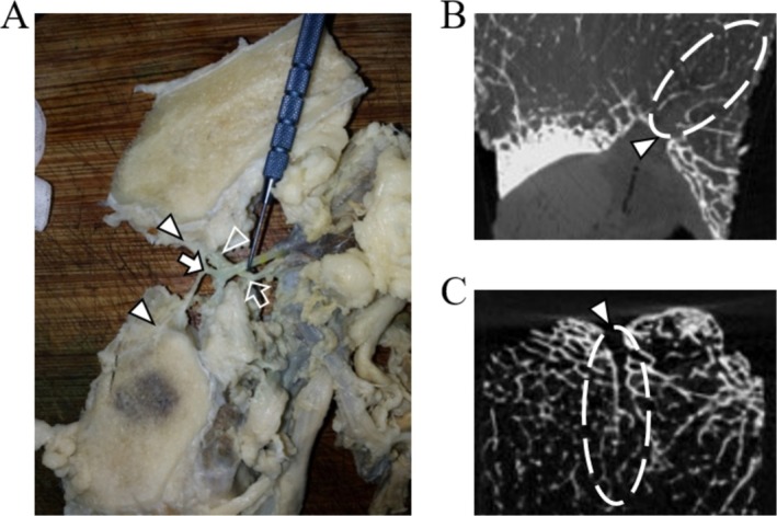
Methods: A newly created composite of MD and Fe3O4 nanoparticles (MD-Fe3O4 NPs) was prepared as a contrast agent to achieve efficacious imaging of the juxta-articular intraosseous vasculature-canal complex (JIVCC). Scanning electron microscopy (SEM) and energy dispersive spectrum (EDS) were employed to observe the structural characteristics and binding stability of the MD-Fe3O4 NPs. In 20 rabbits that received an injection of MD-Fe3O4 NPs, 1-mm-thick computed tomography (CT) scanning was performed for radiographic assessment. Hematoxylin-eosin- and potassium ferrocyanide-stained sections from 10 sacrificed rabbits were used to observe the histological characteristics of JIVCC with MD-Fe3O4 NPs, and the remaining 10 rabbits were utilized for a systemic safety evaluation. After a healthy volunteer received an MD-Fe3O4 NP injection, we also performed CT scanning and related safety evaluations.
Results: When the MD nanoparticles and amino-Fe3O4 nanoparticles were mixed together, they aggregated into a stable compound structure according to microscopic observations and SEM-EDS verification. In 20 rabbits receiving MD-Fe3O4 injections, 1-mm slice CT imaging demonstrated significantly enhanced visualization of the JIVCCs in magnet-placed knees compared to contralateral limbs (tibial JIVCC: p < 0.001; femoral JIVCC: p < 0.001), confirming MD-Fe3O4 NPs as the efficacious magnetic contrast enhancer. The histological characteristics of MD-Fe3O4 NPs in JIVCC were revealed. The levels of serum iron before and 4 and 72 h after MD-Fe3O4 NP injection were 23.9 ± 2.13 μmol/L, 26.2 ± 2.30 μmol/L, and 24.9 ± 2.33 μmol/L, respectively, indicating that there was no significant difference in safety (p = 0.092). After a volunteer received MD-Fe3O4 NPs via intravenous administration, the JIVCC was clearly visualized, laboratory tests of serum iron levels were normal, and no injection-related complications occurred.
Conclusions: A novel compound nanoparticle, which achieved satisfactory overall outcomes, was implemented as an appropriate alternative for the discernible visualization of juxta-articular intraosseous microvessels. The nanotechnology utilized in this study may augment the clinical imaging methodology for the osseous vascular system.
期刊介绍:
Orthopaedic Surgery (OS) is the official journal of the Chinese Orthopaedic Association, focusing on all aspects of orthopaedic technique and surgery.
The journal publishes peer-reviewed articles in the following categories: Original Articles, Clinical Articles, Review Articles, Guidelines, Editorials, Commentaries, Surgical Techniques, Case Reports and Meeting Reports.
 求助内容:
求助内容: 应助结果提醒方式:


