Xinbing Pan, Qianyun Wu, You Zhang, Dongsheng Xu, Xinyuan Zhou, Lianghua Li, Cheng Wang, Weijun Wei, Shuxian An*, Gang Huang* and Jianjun Liu*,
{"title":"结直肠癌cdh17特异性免疫pet显像的发展和临床前评估。","authors":"Xinbing Pan, Qianyun Wu, You Zhang, Dongsheng Xu, Xinyuan Zhou, Lianghua Li, Cheng Wang, Weijun Wei, Shuxian An*, Gang Huang* and Jianjun Liu*, ","doi":"10.1021/acs.molpharmaceut.5c00525","DOIUrl":null,"url":null,"abstract":"<p >Cadherin 17 (CDH17) is found to be abnormally expressed in colorectal cancer (CRC) and linked to a prognosis. Its consistent presence in both primary and metastatic CRC positions it as a promising biomarker. This study aims to develop CDH17-targeted immuno-positron emission tomography (immunoPET) probes and evaluate their potential for diagnosing CRC. Immunohistochemical (IHC) staining was performed on CRC tissue microarrays to analyze CDH17 expression. The CDH17 expression of CRC cells (LS174T, HCT116, and Caco-2) and pancreatic cancer cells (AsPC-1) was detected by flow cytometry. CDH17-specific nanobodies (i.e., CDH1) were produced and labeled with gallium-68 (<sup>68</sup>Ga) and fluorine-18 (<sup>18</sup>F) to generate imaging probes. ImmunoPET imaging was conducted to evaluate the probes’ diagnostic abilities in tumor models. IHC staining of CRC tissue microarrays demonstrated an elevated expression of CDH17 in malignant tissues relative to that in adjacent normal tissues. Flow cytometry showed that CDH17 was expressed on the surface of Caco-2 and AsPC-1 cells but not on the surface of HCT116 or LS174T cells. ImmunoPET imaging with [<sup>68</sup>Ga]Ga-NOTA-CDH1 successfully visualized CDH17 expression in Caco-2 tumors, whereas no significant tracer uptake was observed in HCT116 tumors. [<sup>18</sup>F]AlF-RESCA-CDH1 immunoPET imaging demonstrated CDH17 expression in both LS174T and AsPC-1 tumors. IHC confirmed CDH17 expression on the membrane of Caco-2 and AsPC-1 cells with no significant expression on HCT116 cells. CDH17 expression was evident in the LS174T tumor capsule tissue. In this study, we developed three nanobody-based tracers targeting CDH17, of which [<sup>68</sup>Ga]Ga-NOTA-CDH1 and [<sup>18</sup>F]AlF-RESCA-CDH1 noninvasively demonstrated CDH17 expression in preclinical models.</p>","PeriodicalId":52,"journal":{"name":"Molecular Pharmaceutics","volume":"22 9","pages":"5512–5522"},"PeriodicalIF":4.5000,"publicationDate":"2025-08-10","publicationTypes":"Journal Article","fieldsOfStudy":null,"isOpenAccess":false,"openAccessPdf":"","citationCount":"0","resultStr":"{\"title\":\"Development and Preclinical Evaluation of CDH17-Specific ImmunoPET Imaging in Colorectal Cancers\",\"authors\":\"Xinbing Pan, Qianyun Wu, You Zhang, Dongsheng Xu, Xinyuan Zhou, Lianghua Li, Cheng Wang, Weijun Wei, Shuxian An*, Gang Huang* and Jianjun Liu*, \",\"doi\":\"10.1021/acs.molpharmaceut.5c00525\",\"DOIUrl\":null,\"url\":null,\"abstract\":\"<p >Cadherin 17 (CDH17) is found to be abnormally expressed in colorectal cancer (CRC) and linked to a prognosis. Its consistent presence in both primary and metastatic CRC positions it as a promising biomarker. This study aims to develop CDH17-targeted immuno-positron emission tomography (immunoPET) probes and evaluate their potential for diagnosing CRC. Immunohistochemical (IHC) staining was performed on CRC tissue microarrays to analyze CDH17 expression. The CDH17 expression of CRC cells (LS174T, HCT116, and Caco-2) and pancreatic cancer cells (AsPC-1) was detected by flow cytometry. CDH17-specific nanobodies (i.e., CDH1) were produced and labeled with gallium-68 (<sup>68</sup>Ga) and fluorine-18 (<sup>18</sup>F) to generate imaging probes. ImmunoPET imaging was conducted to evaluate the probes’ diagnostic abilities in tumor models. IHC staining of CRC tissue microarrays demonstrated an elevated expression of CDH17 in malignant tissues relative to that in adjacent normal tissues. Flow cytometry showed that CDH17 was expressed on the surface of Caco-2 and AsPC-1 cells but not on the surface of HCT116 or LS174T cells. ImmunoPET imaging with [<sup>68</sup>Ga]Ga-NOTA-CDH1 successfully visualized CDH17 expression in Caco-2 tumors, whereas no significant tracer uptake was observed in HCT116 tumors. [<sup>18</sup>F]AlF-RESCA-CDH1 immunoPET imaging demonstrated CDH17 expression in both LS174T and AsPC-1 tumors. IHC confirmed CDH17 expression on the membrane of Caco-2 and AsPC-1 cells with no significant expression on HCT116 cells. CDH17 expression was evident in the LS174T tumor capsule tissue. In this study, we developed three nanobody-based tracers targeting CDH17, of which [<sup>68</sup>Ga]Ga-NOTA-CDH1 and [<sup>18</sup>F]AlF-RESCA-CDH1 noninvasively demonstrated CDH17 expression in preclinical models.</p>\",\"PeriodicalId\":52,\"journal\":{\"name\":\"Molecular Pharmaceutics\",\"volume\":\"22 9\",\"pages\":\"5512–5522\"},\"PeriodicalIF\":4.5000,\"publicationDate\":\"2025-08-10\",\"publicationTypes\":\"Journal Article\",\"fieldsOfStudy\":null,\"isOpenAccess\":false,\"openAccessPdf\":\"\",\"citationCount\":\"0\",\"resultStr\":null,\"platform\":\"Semanticscholar\",\"paperid\":null,\"PeriodicalName\":\"Molecular Pharmaceutics\",\"FirstCategoryId\":\"3\",\"ListUrlMain\":\"https://pubs.acs.org/doi/10.1021/acs.molpharmaceut.5c00525\",\"RegionNum\":2,\"RegionCategory\":\"医学\",\"ArticlePicture\":[],\"TitleCN\":null,\"AbstractTextCN\":null,\"PMCID\":null,\"EPubDate\":\"\",\"PubModel\":\"\",\"JCR\":\"Q2\",\"JCRName\":\"MEDICINE, RESEARCH & EXPERIMENTAL\",\"Score\":null,\"Total\":0}","platform":"Semanticscholar","paperid":null,"PeriodicalName":"Molecular Pharmaceutics","FirstCategoryId":"3","ListUrlMain":"https://pubs.acs.org/doi/10.1021/acs.molpharmaceut.5c00525","RegionNum":2,"RegionCategory":"医学","ArticlePicture":[],"TitleCN":null,"AbstractTextCN":null,"PMCID":null,"EPubDate":"","PubModel":"","JCR":"Q2","JCRName":"MEDICINE, RESEARCH & EXPERIMENTAL","Score":null,"Total":0}
Development and Preclinical Evaluation of CDH17-Specific ImmunoPET Imaging in Colorectal Cancers
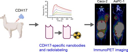
Cadherin 17 (CDH17) is found to be abnormally expressed in colorectal cancer (CRC) and linked to a prognosis. Its consistent presence in both primary and metastatic CRC positions it as a promising biomarker. This study aims to develop CDH17-targeted immuno-positron emission tomography (immunoPET) probes and evaluate their potential for diagnosing CRC. Immunohistochemical (IHC) staining was performed on CRC tissue microarrays to analyze CDH17 expression. The CDH17 expression of CRC cells (LS174T, HCT116, and Caco-2) and pancreatic cancer cells (AsPC-1) was detected by flow cytometry. CDH17-specific nanobodies (i.e., CDH1) were produced and labeled with gallium-68 (68Ga) and fluorine-18 (18F) to generate imaging probes. ImmunoPET imaging was conducted to evaluate the probes’ diagnostic abilities in tumor models. IHC staining of CRC tissue microarrays demonstrated an elevated expression of CDH17 in malignant tissues relative to that in adjacent normal tissues. Flow cytometry showed that CDH17 was expressed on the surface of Caco-2 and AsPC-1 cells but not on the surface of HCT116 or LS174T cells. ImmunoPET imaging with [68Ga]Ga-NOTA-CDH1 successfully visualized CDH17 expression in Caco-2 tumors, whereas no significant tracer uptake was observed in HCT116 tumors. [18F]AlF-RESCA-CDH1 immunoPET imaging demonstrated CDH17 expression in both LS174T and AsPC-1 tumors. IHC confirmed CDH17 expression on the membrane of Caco-2 and AsPC-1 cells with no significant expression on HCT116 cells. CDH17 expression was evident in the LS174T tumor capsule tissue. In this study, we developed three nanobody-based tracers targeting CDH17, of which [68Ga]Ga-NOTA-CDH1 and [18F]AlF-RESCA-CDH1 noninvasively demonstrated CDH17 expression in preclinical models.
期刊介绍:
Molecular Pharmaceutics publishes the results of original research that contributes significantly to the molecular mechanistic understanding of drug delivery and drug delivery systems. The journal encourages contributions describing research at the interface of drug discovery and drug development.
Scientific areas within the scope of the journal include physical and pharmaceutical chemistry, biochemistry and biophysics, molecular and cellular biology, and polymer and materials science as they relate to drug and drug delivery system efficacy. Mechanistic Drug Delivery and Drug Targeting research on modulating activity and efficacy of a drug or drug product is within the scope of Molecular Pharmaceutics. Theoretical and experimental peer-reviewed research articles, communications, reviews, and perspectives are welcomed.
 求助内容:
求助内容: 应助结果提醒方式:


