Melanie A Reuter, Rosalinda Moreno, Madelynn E Agabao-Tucker, Rahaf Shishani, Jessica Miranda Bustamante, Zara Marfori, Taylor Richieri, Anthony E Valenzuela, Ameer Y Taha, Pamela J Lein, Renu Nandakumar, Bethany P Cummings
{"title":"啮齿动物脑区胆汁酸谱不同,在阿尔茨海默病啮齿动物模型中被破坏。","authors":"Melanie A Reuter, Rosalinda Moreno, Madelynn E Agabao-Tucker, Rahaf Shishani, Jessica Miranda Bustamante, Zara Marfori, Taylor Richieri, Anthony E Valenzuela, Ameer Y Taha, Pamela J Lein, Renu Nandakumar, Bethany P Cummings","doi":"10.1002/cph4.70034","DOIUrl":null,"url":null,"abstract":"<p><p>Low but biologically relevant levels of bile acids are found in the brain and are altered in patients with Alzheimer's disease (AD). However, the regulation of brain bile acid levels and what drives brain bile acid dynamics are poorly understood. Bile acids are synthesized in the liver and further metabolized by bacteria in the gut. Therefore, bile acids are mediators of the liver-brain axis and the gut-brain axis. Additionally, whether the bile acid profile differs between brain regions and whether the brain region-specific bile acid profile is impacted by disease, such as AD, is unknown. Therefore, we tested the hypothesis that the brain bile acid profile is influenced by peripheral bile acid metabolism, differs between brain regions, and that these dynamics change in AD. To this end, we assessed the bile acid profile in the cortex and hippocampus of wild-type mice maintained on different diets. To test the effect of AD, we used the TgF344-AD rat model. We found that the brain bile acid profile in mice was mildly altered by diet and, in both mice and rats, differs substantially between brain regions. For example, cholic acid and taurocholic acid are enriched in the cortex relative to the hippocampus in both mice and rats. Further, using a rat model of AD, we found that brain region differences in bile acid profiles are attenuated in AD. Together, these data demonstrate that both peripheral and central regulatory mechanisms maintain bile acid homeostasis in specific brain regions and that these homeostatic mechanisms are disrupted in AD.</p>","PeriodicalId":10573,"journal":{"name":"Comprehensive Physiology","volume":"15 4","pages":"e70034"},"PeriodicalIF":5.2000,"publicationDate":"2025-08-01","publicationTypes":"Journal Article","fieldsOfStudy":null,"isOpenAccess":false,"openAccessPdf":"https://www.ncbi.nlm.nih.gov/pmc/articles/PMC12320573/pdf/","citationCount":"0","resultStr":"{\"title\":\"Bile Acid Profile Differs Between Brain Regions in Rodents and Is Disrupted in a Rodent Model of Alzheimer's Disease.\",\"authors\":\"Melanie A Reuter, Rosalinda Moreno, Madelynn E Agabao-Tucker, Rahaf Shishani, Jessica Miranda Bustamante, Zara Marfori, Taylor Richieri, Anthony E Valenzuela, Ameer Y Taha, Pamela J Lein, Renu Nandakumar, Bethany P Cummings\",\"doi\":\"10.1002/cph4.70034\",\"DOIUrl\":null,\"url\":null,\"abstract\":\"<p><p>Low but biologically relevant levels of bile acids are found in the brain and are altered in patients with Alzheimer's disease (AD). However, the regulation of brain bile acid levels and what drives brain bile acid dynamics are poorly understood. Bile acids are synthesized in the liver and further metabolized by bacteria in the gut. Therefore, bile acids are mediators of the liver-brain axis and the gut-brain axis. Additionally, whether the bile acid profile differs between brain regions and whether the brain region-specific bile acid profile is impacted by disease, such as AD, is unknown. Therefore, we tested the hypothesis that the brain bile acid profile is influenced by peripheral bile acid metabolism, differs between brain regions, and that these dynamics change in AD. To this end, we assessed the bile acid profile in the cortex and hippocampus of wild-type mice maintained on different diets. To test the effect of AD, we used the TgF344-AD rat model. We found that the brain bile acid profile in mice was mildly altered by diet and, in both mice and rats, differs substantially between brain regions. For example, cholic acid and taurocholic acid are enriched in the cortex relative to the hippocampus in both mice and rats. Further, using a rat model of AD, we found that brain region differences in bile acid profiles are attenuated in AD. Together, these data demonstrate that both peripheral and central regulatory mechanisms maintain bile acid homeostasis in specific brain regions and that these homeostatic mechanisms are disrupted in AD.</p>\",\"PeriodicalId\":10573,\"journal\":{\"name\":\"Comprehensive Physiology\",\"volume\":\"15 4\",\"pages\":\"e70034\"},\"PeriodicalIF\":5.2000,\"publicationDate\":\"2025-08-01\",\"publicationTypes\":\"Journal Article\",\"fieldsOfStudy\":null,\"isOpenAccess\":false,\"openAccessPdf\":\"https://www.ncbi.nlm.nih.gov/pmc/articles/PMC12320573/pdf/\",\"citationCount\":\"0\",\"resultStr\":null,\"platform\":\"Semanticscholar\",\"paperid\":null,\"PeriodicalName\":\"Comprehensive Physiology\",\"FirstCategoryId\":\"3\",\"ListUrlMain\":\"https://doi.org/10.1002/cph4.70034\",\"RegionNum\":2,\"RegionCategory\":\"医学\",\"ArticlePicture\":[],\"TitleCN\":null,\"AbstractTextCN\":null,\"PMCID\":null,\"EPubDate\":\"\",\"PubModel\":\"\",\"JCR\":\"Q1\",\"JCRName\":\"PHYSIOLOGY\",\"Score\":null,\"Total\":0}","platform":"Semanticscholar","paperid":null,"PeriodicalName":"Comprehensive Physiology","FirstCategoryId":"3","ListUrlMain":"https://doi.org/10.1002/cph4.70034","RegionNum":2,"RegionCategory":"医学","ArticlePicture":[],"TitleCN":null,"AbstractTextCN":null,"PMCID":null,"EPubDate":"","PubModel":"","JCR":"Q1","JCRName":"PHYSIOLOGY","Score":null,"Total":0}
Bile Acid Profile Differs Between Brain Regions in Rodents and Is Disrupted in a Rodent Model of Alzheimer's Disease.
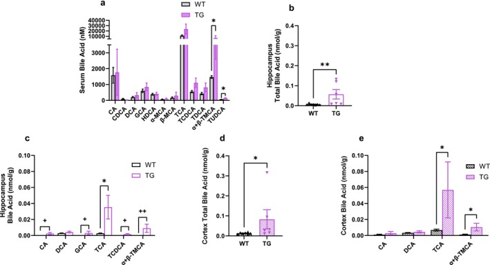
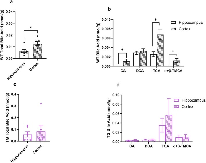
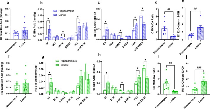
Low but biologically relevant levels of bile acids are found in the brain and are altered in patients with Alzheimer's disease (AD). However, the regulation of brain bile acid levels and what drives brain bile acid dynamics are poorly understood. Bile acids are synthesized in the liver and further metabolized by bacteria in the gut. Therefore, bile acids are mediators of the liver-brain axis and the gut-brain axis. Additionally, whether the bile acid profile differs between brain regions and whether the brain region-specific bile acid profile is impacted by disease, such as AD, is unknown. Therefore, we tested the hypothesis that the brain bile acid profile is influenced by peripheral bile acid metabolism, differs between brain regions, and that these dynamics change in AD. To this end, we assessed the bile acid profile in the cortex and hippocampus of wild-type mice maintained on different diets. To test the effect of AD, we used the TgF344-AD rat model. We found that the brain bile acid profile in mice was mildly altered by diet and, in both mice and rats, differs substantially between brain regions. For example, cholic acid and taurocholic acid are enriched in the cortex relative to the hippocampus in both mice and rats. Further, using a rat model of AD, we found that brain region differences in bile acid profiles are attenuated in AD. Together, these data demonstrate that both peripheral and central regulatory mechanisms maintain bile acid homeostasis in specific brain regions and that these homeostatic mechanisms are disrupted in AD.
期刊介绍:
Comprehensive Physiology is the most authoritative and comprehensive collection of physiology information ever assembled, and uses the most powerful features of review journals and electronic reference works to cover the latest key developments in the field, through the most authoritative articles on the subjects covered.
This makes Comprehensive Physiology a valued reference work on the evolving science of physiology for both researchers and clinicians. It also provides a useful teaching tool for instructors and an informative resource for medical students and other students in the life and health sciences.

 求助内容:
求助内容: 应助结果提醒方式:


