Norsyuhadah Musa, Wan Norlina Wan Azman, Nor Amirah Mohammad Nazri, Tuan Salwani Tuan Ismail, Azian Harun, Najib Majdi Yaacob, Sarina Sulong, K N S Sirajudeen, Mahaya Che Mat, Hani Ajrina Zulkeflee
{"title":"COVID-19患者α -1抗胰蛋白酶:马来西亚双中心筛查研究","authors":"Norsyuhadah Musa, Wan Norlina Wan Azman, Nor Amirah Mohammad Nazri, Tuan Salwani Tuan Ismail, Azian Harun, Najib Majdi Yaacob, Sarina Sulong, K N S Sirajudeen, Mahaya Che Mat, Hani Ajrina Zulkeflee","doi":"10.5144/0256-4947.2025.225","DOIUrl":null,"url":null,"abstract":"<p><strong>Background: </strong>Alpha-1 antitrypsin (A1AT) deficiency has been recognized as an adverse prognostic determinant in severe instances of COVID-19.</p><p><strong>Objective: </strong>To determine the A1AT phenotypes and levels in individuals at various clinical stages of COVID-19 compared to healthy controls.</p><p><strong>Design: </strong>Case-control study.</p><p><strong>Settings: </strong>Hospital Raja Perempuan Zainab II (HRPZ II) and Hospital Ampang, Malaysia.</p><p><strong>Patients and methods: </strong>The analysis included a total of 282 patients. We categorized 188 COVID-19 patients from two centres in Malaysia into two groups: mild to moderate (stages 1-3) and severe to critical (stages 4-5) and compared them with 94 healthy controls.</p><p><strong>Main outcome measures: </strong>A1AT phenotypes and levels in different COVID-19 stages compared to healthy controls.</p><p><strong>Sample size: </strong>282 subjects.</p><p><strong>Results: </strong>The frequency (n) and percentage (%) in the control group, 88 (93.6) exhibited PiMM phenotypes, whereas 6 (6.4) displayed PiXM/PiYM phenotypes. Within the mild to moderate COVID-19 group, 88 (93.6) had PiMM phenotypes, 3 (3.2) featured PiXM/PiYM, and 1 presented PiBM phenotypes. Among severe to critical COVID-19 patients, the PiMM phenotype was identified in 61 (64.9) with 16 (17) having PiBM phenotypes, 4 (4.5) displaying PiCM, 2 (2.1) featuring PiXM/PiYM, and 1 (1.1) presenting PiEM phenotypes. Variants such as MS, MZ, S, and Z were undetected. However, 12 COVID-19 patient samples yielded inconclusive results. Median (IQR: 25th to 75th percentile) A1AT concentrations for controls were 1.8 (1.3-2.3) g/L, for mild to moderate cases 1.9 (1.2-2.6) g/L, and for severe to critical COVID-19 cases 2.1 (1.4-2.8) g/L.</p><p><strong>Conclusion: </strong>This research identifies the PiMM phenotype as the predominant phenotype expression within the studied population. This prevalence underscores the potential role of genetic factors in determining the biological response to SARS-CoV-2 infection. The presence of another phenotype variant across the study population suggests a nuanced genetic landscape that warrants further exploration.</p><p><strong>Limitation: </strong>The absence of follow-up A1AT quantification and baseline measurements limits the assessment of disease progression. The isolectric focusing phenotyping technique used might have missed specific A1ATD variants.</p>","PeriodicalId":93875,"journal":{"name":"Annals of Saudi medicine","volume":"45 4","pages":"225-234"},"PeriodicalIF":0.0000,"publicationDate":"2025-07-01","publicationTypes":"Journal Article","fieldsOfStudy":null,"isOpenAccess":false,"openAccessPdf":"https://www.ncbi.nlm.nih.gov/pmc/articles/PMC12318249/pdf/","citationCount":"0","resultStr":"{\"title\":\"Alpha-1 antitrypsin in COVID-19 patients: a dual-center screening study in Malaysia.\",\"authors\":\"Norsyuhadah Musa, Wan Norlina Wan Azman, Nor Amirah Mohammad Nazri, Tuan Salwani Tuan Ismail, Azian Harun, Najib Majdi Yaacob, Sarina Sulong, K N S Sirajudeen, Mahaya Che Mat, Hani Ajrina Zulkeflee\",\"doi\":\"10.5144/0256-4947.2025.225\",\"DOIUrl\":null,\"url\":null,\"abstract\":\"<p><strong>Background: </strong>Alpha-1 antitrypsin (A1AT) deficiency has been recognized as an adverse prognostic determinant in severe instances of COVID-19.</p><p><strong>Objective: </strong>To determine the A1AT phenotypes and levels in individuals at various clinical stages of COVID-19 compared to healthy controls.</p><p><strong>Design: </strong>Case-control study.</p><p><strong>Settings: </strong>Hospital Raja Perempuan Zainab II (HRPZ II) and Hospital Ampang, Malaysia.</p><p><strong>Patients and methods: </strong>The analysis included a total of 282 patients. We categorized 188 COVID-19 patients from two centres in Malaysia into two groups: mild to moderate (stages 1-3) and severe to critical (stages 4-5) and compared them with 94 healthy controls.</p><p><strong>Main outcome measures: </strong>A1AT phenotypes and levels in different COVID-19 stages compared to healthy controls.</p><p><strong>Sample size: </strong>282 subjects.</p><p><strong>Results: </strong>The frequency (n) and percentage (%) in the control group, 88 (93.6) exhibited PiMM phenotypes, whereas 6 (6.4) displayed PiXM/PiYM phenotypes. Within the mild to moderate COVID-19 group, 88 (93.6) had PiMM phenotypes, 3 (3.2) featured PiXM/PiYM, and 1 presented PiBM phenotypes. Among severe to critical COVID-19 patients, the PiMM phenotype was identified in 61 (64.9) with 16 (17) having PiBM phenotypes, 4 (4.5) displaying PiCM, 2 (2.1) featuring PiXM/PiYM, and 1 (1.1) presenting PiEM phenotypes. Variants such as MS, MZ, S, and Z were undetected. However, 12 COVID-19 patient samples yielded inconclusive results. Median (IQR: 25th to 75th percentile) A1AT concentrations for controls were 1.8 (1.3-2.3) g/L, for mild to moderate cases 1.9 (1.2-2.6) g/L, and for severe to critical COVID-19 cases 2.1 (1.4-2.8) g/L.</p><p><strong>Conclusion: </strong>This research identifies the PiMM phenotype as the predominant phenotype expression within the studied population. This prevalence underscores the potential role of genetic factors in determining the biological response to SARS-CoV-2 infection. The presence of another phenotype variant across the study population suggests a nuanced genetic landscape that warrants further exploration.</p><p><strong>Limitation: </strong>The absence of follow-up A1AT quantification and baseline measurements limits the assessment of disease progression. The isolectric focusing phenotyping technique used might have missed specific A1ATD variants.</p>\",\"PeriodicalId\":93875,\"journal\":{\"name\":\"Annals of Saudi medicine\",\"volume\":\"45 4\",\"pages\":\"225-234\"},\"PeriodicalIF\":0.0000,\"publicationDate\":\"2025-07-01\",\"publicationTypes\":\"Journal Article\",\"fieldsOfStudy\":null,\"isOpenAccess\":false,\"openAccessPdf\":\"https://www.ncbi.nlm.nih.gov/pmc/articles/PMC12318249/pdf/\",\"citationCount\":\"0\",\"resultStr\":null,\"platform\":\"Semanticscholar\",\"paperid\":null,\"PeriodicalName\":\"Annals of Saudi medicine\",\"FirstCategoryId\":\"1085\",\"ListUrlMain\":\"https://doi.org/10.5144/0256-4947.2025.225\",\"RegionNum\":0,\"RegionCategory\":null,\"ArticlePicture\":[],\"TitleCN\":null,\"AbstractTextCN\":null,\"PMCID\":null,\"EPubDate\":\"2025/8/7 0:00:00\",\"PubModel\":\"Epub\",\"JCR\":\"\",\"JCRName\":\"\",\"Score\":null,\"Total\":0}","platform":"Semanticscholar","paperid":null,"PeriodicalName":"Annals of Saudi medicine","FirstCategoryId":"1085","ListUrlMain":"https://doi.org/10.5144/0256-4947.2025.225","RegionNum":0,"RegionCategory":null,"ArticlePicture":[],"TitleCN":null,"AbstractTextCN":null,"PMCID":null,"EPubDate":"2025/8/7 0:00:00","PubModel":"Epub","JCR":"","JCRName":"","Score":null,"Total":0}
Alpha-1 antitrypsin in COVID-19 patients: a dual-center screening study in Malaysia.
Background: Alpha-1 antitrypsin (A1AT) deficiency has been recognized as an adverse prognostic determinant in severe instances of COVID-19.
Objective: To determine the A1AT phenotypes and levels in individuals at various clinical stages of COVID-19 compared to healthy controls.
Design: Case-control study.
Settings: Hospital Raja Perempuan Zainab II (HRPZ II) and Hospital Ampang, Malaysia.
Patients and methods: The analysis included a total of 282 patients. We categorized 188 COVID-19 patients from two centres in Malaysia into two groups: mild to moderate (stages 1-3) and severe to critical (stages 4-5) and compared them with 94 healthy controls.
Main outcome measures: A1AT phenotypes and levels in different COVID-19 stages compared to healthy controls.
Sample size: 282 subjects.
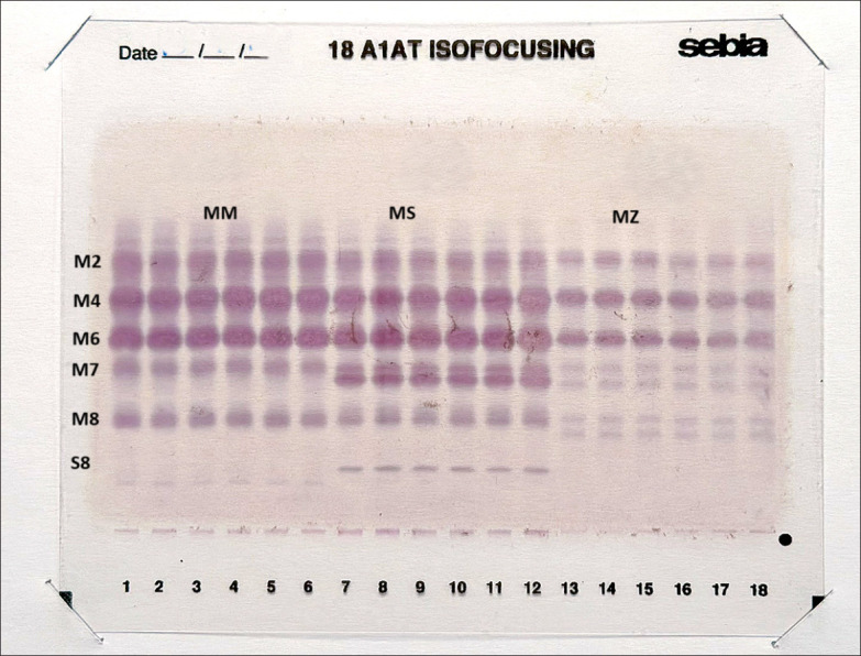
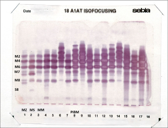
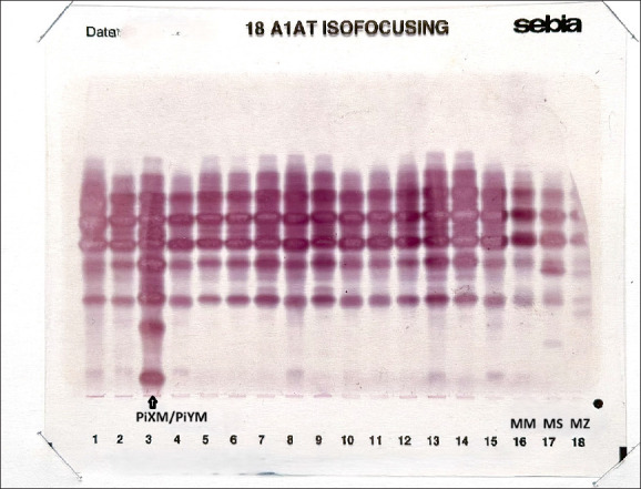
Results: The frequency (n) and percentage (%) in the control group, 88 (93.6) exhibited PiMM phenotypes, whereas 6 (6.4) displayed PiXM/PiYM phenotypes. Within the mild to moderate COVID-19 group, 88 (93.6) had PiMM phenotypes, 3 (3.2) featured PiXM/PiYM, and 1 presented PiBM phenotypes. Among severe to critical COVID-19 patients, the PiMM phenotype was identified in 61 (64.9) with 16 (17) having PiBM phenotypes, 4 (4.5) displaying PiCM, 2 (2.1) featuring PiXM/PiYM, and 1 (1.1) presenting PiEM phenotypes. Variants such as MS, MZ, S, and Z were undetected. However, 12 COVID-19 patient samples yielded inconclusive results. Median (IQR: 25th to 75th percentile) A1AT concentrations for controls were 1.8 (1.3-2.3) g/L, for mild to moderate cases 1.9 (1.2-2.6) g/L, and for severe to critical COVID-19 cases 2.1 (1.4-2.8) g/L.
Conclusion: This research identifies the PiMM phenotype as the predominant phenotype expression within the studied population. This prevalence underscores the potential role of genetic factors in determining the biological response to SARS-CoV-2 infection. The presence of another phenotype variant across the study population suggests a nuanced genetic landscape that warrants further exploration.
Limitation: The absence of follow-up A1AT quantification and baseline measurements limits the assessment of disease progression. The isolectric focusing phenotyping technique used might have missed specific A1ATD variants.

 求助内容:
求助内容: 应助结果提醒方式:


