三维低剂量、少视点心脏SPECT成像的可推广扩散框架。
IF 11.8
1区 医学
Q1 COMPUTER SCIENCE, ARTIFICIAL INTELLIGENCE
引用次数: 0
摘要
SPECT心肌灌注成像被广泛用于冠状动脉疾病的诊断,但在低剂量和少视点采集环境下,图像质量会受到负面影响。虽然已经引入了各种深度学习方法来提高低剂量或少视点SPECT数据的图像质量,但以前的方法往往不能推广到不同的采集设置,限制了实际的适用性。这项工作引入了DiffSPECT-3D,这是一种用于心脏3D SPECT成像的扩散框架,可以有效地适应不同的采集设置,而无需进一步的网络重新训练或微调。利用图像和投影数据,提出了一种一致性策略,以确保每一步的扩散采样与低剂量/少视图投影测量值、图像数据和扫描仪几何形状一致,从而能够推广到不同的低剂量/少视图设置。结合CT的解剖空间信息和总变异约束,我们提出了一个2.5D条件策略,允许DiffSPECT-3D从整个图像体中观察3D上下文信息,解决了扩散模型中的3D记忆/计算问题。我们对来自795名患者的1325项临床99mTc四氟磷应激/休息研究进行了广泛的评估。将每项研究重构为5个不同的低计数水平和5个不同的投影少视图水平,分别从1%到50%和1到9视图进行模型评估。与心导管检查结果和核心脏病专家的诊断回顾相对照,本文的结果显示了在不影响临床表现的情况下实现低剂量和少视点SPECT成像的潜力。此外,DiffSPECT-3D可以直接应用于全剂量SPECT图像,以进一步提高图像质量,特别是在低剂量应力优先的心脏SPECT成像方案中。本文章由计算机程序翻译,如有差异,请以英文原文为准。

A generalizable diffusion framework for 3D low-dose and few-view cardiac SPECT imaging
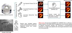
Myocardial perfusion imaging using SPECT is widely utilized to diagnose coronary artery diseases, but image quality can be negatively affected in low-dose and few-view acquisition settings. Although various deep learning methods have been introduced to improve image quality from low-dose or few-view SPECT data, previous approaches often fail to generalize across different acquisition settings, limiting realistic applicability. This work introduced DiffSPECT-3D, a diffusion framework for 3D cardiac SPECT imaging that effectively adapts to different acquisition settings without requiring further network re-training or fine-tuning. Using both image and projection data, a consistency strategy is proposed to ensure that diffusion sampling at each step aligns with the low-dose/few-view projection measurements, the image data, and the scanner geometry, thus enabling generalization to different low-dose/few-view settings. Incorporating anatomical spatial information from CT and total variation constraint, we proposed a 2.5D conditional strategy to allow DiffSPECT-3D to observe 3D contextual information from the entire image volume, addressing the 3D memory/computational issues in diffusion model. We extensively evaluated the proposed method on 1,325 clinical Tc tetrofosmin stress/rest studies from 795 patients. Each study was reconstructed into 5 different low-count levels and 5 different projection few-view levels for model evaluations, ranging from 1% to 50% and from 1 view to 9 view, respectively. Validated against cardiac catheterization results and diagnostic review from nuclear cardiologists, the presented results show the potential to achieve low-dose and few-view SPECT imaging without compromising clinical performance. Additionally, DiffSPECT-3D could be directly applied to full-dose SPECT images to further improve image quality, especially in a low-dose stress-first cardiac SPECT imaging protocol.
求助全文
通过发布文献求助,成功后即可免费获取论文全文。
去求助
来源期刊

Medical image analysis
工程技术-工程:生物医学
CiteScore
22.10
自引率
6.40%
发文量
309
审稿时长
6.6 months
期刊介绍:
Medical Image Analysis serves as a platform for sharing new research findings in the realm of medical and biological image analysis, with a focus on applications of computer vision, virtual reality, and robotics to biomedical imaging challenges. The journal prioritizes the publication of high-quality, original papers contributing to the fundamental science of processing, analyzing, and utilizing medical and biological images. It welcomes approaches utilizing biomedical image datasets across all spatial scales, from molecular/cellular imaging to tissue/organ imaging.
 求助内容:
求助内容: 应助结果提醒方式:


