Maxwell C. Campbell, Steven I. Pollmann, Jaques S. Milner, Emily A. Lalone, David W. Holdsworth
{"title":"一种模拟3D打印物体射线照片的新方法","authors":"Maxwell C. Campbell, Steven I. Pollmann, Jaques S. Milner, Emily A. Lalone, David W. Holdsworth","doi":"10.1002/acm2.70159","DOIUrl":null,"url":null,"abstract":"<div>\n \n \n <section>\n \n <h3> Background</h3>\n \n <p>3D printing has a number of applications within medicine and healthcare. In applications involving radiography, the internal infill structure and external geometry of a 3D printed part can produce undesirable artifacts, limiting the full potential of 3D printing as a manufacturing technology. While the mechanical performance of a 3D printed part can be easily simulated, it is difficult to simulate the radiographic artifact produced.</p>\n </section>\n \n <section>\n \n <h3> Purpose</h3>\n \n <p>The purpose of this work was to develop a tool that allows users to simulate the radiographic artifact produced by a 3D printed object.</p>\n </section>\n \n <section>\n \n <h3> Methods</h3>\n \n <p>Three regular hexagons of identical geometry were sliced and 3D printed using polylactic acid (PLA) filament on a fused deposition modeling (FDM) 3D printer with varying infill patterns: rectilinear grid, cubic, and gyroid. The hexagons were then radiographed using clinical-standard scanning protocols. The captured radiographs were compared to simulated radiographs generated using the G-Code developed when the objects were sliced. The physical and simulated virtual radiographs were compared to one another, and the simulated angle of least and greatest artifact was noted.</p>\n </section>\n \n <section>\n \n <h3> Results</h3>\n \n <p>Strong visual agreement was found between the physically captured and simulated virtual radiographs. The projection angles that produced the least amount of artifact were 22.5°, 22.5°, and 12.25° for grid, cubic, and gyroid infills, respectively. The projection angles that produced the greatest amount of artifact were 0°, 45°, and 45° for grid, cubic, and gyroid infills, respectively.</p>\n </section>\n \n <section>\n \n <h3> Conclusions</h3>\n \n <p>This work provides designers of 3D printed components with a new way to evaluate a design's radiographic performance. Previously, designers would have to physically print and radiograph a part to determine the artifact produced. This work outlines the development of a tool that simulates the radiograph of a 3D printed part from multiple different projections, saving designers time to iterate to their final design.</p>\n </section>\n </div>","PeriodicalId":14989,"journal":{"name":"Journal of Applied Clinical Medical Physics","volume":"26 8","pages":""},"PeriodicalIF":2.2000,"publicationDate":"2025-08-03","publicationTypes":"Journal Article","fieldsOfStudy":null,"isOpenAccess":false,"openAccessPdf":"https://onlinelibrary.wiley.com/doi/epdf/10.1002/acm2.70159","citationCount":"0","resultStr":"{\"title\":\"A novel method to simulate radiographs of 3D printed objects\",\"authors\":\"Maxwell C. Campbell, Steven I. Pollmann, Jaques S. Milner, Emily A. Lalone, David W. Holdsworth\",\"doi\":\"10.1002/acm2.70159\",\"DOIUrl\":null,\"url\":null,\"abstract\":\"<div>\\n \\n \\n <section>\\n \\n <h3> Background</h3>\\n \\n <p>3D printing has a number of applications within medicine and healthcare. In applications involving radiography, the internal infill structure and external geometry of a 3D printed part can produce undesirable artifacts, limiting the full potential of 3D printing as a manufacturing technology. While the mechanical performance of a 3D printed part can be easily simulated, it is difficult to simulate the radiographic artifact produced.</p>\\n </section>\\n \\n <section>\\n \\n <h3> Purpose</h3>\\n \\n <p>The purpose of this work was to develop a tool that allows users to simulate the radiographic artifact produced by a 3D printed object.</p>\\n </section>\\n \\n <section>\\n \\n <h3> Methods</h3>\\n \\n <p>Three regular hexagons of identical geometry were sliced and 3D printed using polylactic acid (PLA) filament on a fused deposition modeling (FDM) 3D printer with varying infill patterns: rectilinear grid, cubic, and gyroid. The hexagons were then radiographed using clinical-standard scanning protocols. The captured radiographs were compared to simulated radiographs generated using the G-Code developed when the objects were sliced. The physical and simulated virtual radiographs were compared to one another, and the simulated angle of least and greatest artifact was noted.</p>\\n </section>\\n \\n <section>\\n \\n <h3> Results</h3>\\n \\n <p>Strong visual agreement was found between the physically captured and simulated virtual radiographs. The projection angles that produced the least amount of artifact were 22.5°, 22.5°, and 12.25° for grid, cubic, and gyroid infills, respectively. The projection angles that produced the greatest amount of artifact were 0°, 45°, and 45° for grid, cubic, and gyroid infills, respectively.</p>\\n </section>\\n \\n <section>\\n \\n <h3> Conclusions</h3>\\n \\n <p>This work provides designers of 3D printed components with a new way to evaluate a design's radiographic performance. Previously, designers would have to physically print and radiograph a part to determine the artifact produced. This work outlines the development of a tool that simulates the radiograph of a 3D printed part from multiple different projections, saving designers time to iterate to their final design.</p>\\n </section>\\n </div>\",\"PeriodicalId\":14989,\"journal\":{\"name\":\"Journal of Applied Clinical Medical Physics\",\"volume\":\"26 8\",\"pages\":\"\"},\"PeriodicalIF\":2.2000,\"publicationDate\":\"2025-08-03\",\"publicationTypes\":\"Journal Article\",\"fieldsOfStudy\":null,\"isOpenAccess\":false,\"openAccessPdf\":\"https://onlinelibrary.wiley.com/doi/epdf/10.1002/acm2.70159\",\"citationCount\":\"0\",\"resultStr\":null,\"platform\":\"Semanticscholar\",\"paperid\":null,\"PeriodicalName\":\"Journal of Applied Clinical Medical Physics\",\"FirstCategoryId\":\"3\",\"ListUrlMain\":\"https://aapm.onlinelibrary.wiley.com/doi/10.1002/acm2.70159\",\"RegionNum\":4,\"RegionCategory\":\"医学\",\"ArticlePicture\":[],\"TitleCN\":null,\"AbstractTextCN\":null,\"PMCID\":null,\"EPubDate\":\"\",\"PubModel\":\"\",\"JCR\":\"Q3\",\"JCRName\":\"RADIOLOGY, NUCLEAR MEDICINE & MEDICAL IMAGING\",\"Score\":null,\"Total\":0}","platform":"Semanticscholar","paperid":null,"PeriodicalName":"Journal of Applied Clinical Medical Physics","FirstCategoryId":"3","ListUrlMain":"https://aapm.onlinelibrary.wiley.com/doi/10.1002/acm2.70159","RegionNum":4,"RegionCategory":"医学","ArticlePicture":[],"TitleCN":null,"AbstractTextCN":null,"PMCID":null,"EPubDate":"","PubModel":"","JCR":"Q3","JCRName":"RADIOLOGY, NUCLEAR MEDICINE & MEDICAL IMAGING","Score":null,"Total":0}
A novel method to simulate radiographs of 3D printed objects
Background
3D printing has a number of applications within medicine and healthcare. In applications involving radiography, the internal infill structure and external geometry of a 3D printed part can produce undesirable artifacts, limiting the full potential of 3D printing as a manufacturing technology. While the mechanical performance of a 3D printed part can be easily simulated, it is difficult to simulate the radiographic artifact produced.
Purpose
The purpose of this work was to develop a tool that allows users to simulate the radiographic artifact produced by a 3D printed object.
Methods
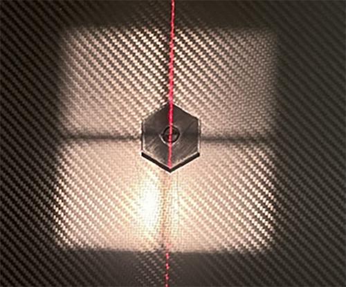
Three regular hexagons of identical geometry were sliced and 3D printed using polylactic acid (PLA) filament on a fused deposition modeling (FDM) 3D printer with varying infill patterns: rectilinear grid, cubic, and gyroid. The hexagons were then radiographed using clinical-standard scanning protocols. The captured radiographs were compared to simulated radiographs generated using the G-Code developed when the objects were sliced. The physical and simulated virtual radiographs were compared to one another, and the simulated angle of least and greatest artifact was noted.
Results
Strong visual agreement was found between the physically captured and simulated virtual radiographs. The projection angles that produced the least amount of artifact were 22.5°, 22.5°, and 12.25° for grid, cubic, and gyroid infills, respectively. The projection angles that produced the greatest amount of artifact were 0°, 45°, and 45° for grid, cubic, and gyroid infills, respectively.
Conclusions
This work provides designers of 3D printed components with a new way to evaluate a design's radiographic performance. Previously, designers would have to physically print and radiograph a part to determine the artifact produced. This work outlines the development of a tool that simulates the radiograph of a 3D printed part from multiple different projections, saving designers time to iterate to their final design.
期刊介绍:
Journal of Applied Clinical Medical Physics is an international Open Access publication dedicated to clinical medical physics. JACMP welcomes original contributions dealing with all aspects of medical physics from scientists working in the clinical medical physics around the world. JACMP accepts only online submission.
JACMP will publish:
-Original Contributions: Peer-reviewed, investigations that represent new and significant contributions to the field. Recommended word count: up to 7500.
-Review Articles: Reviews of major areas or sub-areas in the field of clinical medical physics. These articles may be of any length and are peer reviewed.
-Technical Notes: These should be no longer than 3000 words, including key references.
-Letters to the Editor: Comments on papers published in JACMP or on any other matters of interest to clinical medical physics. These should not be more than 1250 (including the literature) and their publication is only based on the decision of the editor, who occasionally asks experts on the merit of the contents.
-Book Reviews: The editorial office solicits Book Reviews.
-Announcements of Forthcoming Meetings: The Editor may provide notice of forthcoming meetings, course offerings, and other events relevant to clinical medical physics.
-Parallel Opposed Editorial: We welcome topics relevant to clinical practice and medical physics profession. The contents can be controversial debate or opposed aspects of an issue. One author argues for the position and the other against. Each side of the debate contains an opening statement up to 800 words, followed by a rebuttal up to 500 words. Readers interested in participating in this series should contact the moderator with a proposed title and a short description of the topic


 求助内容:
求助内容: 应助结果提醒方式:


