Thuy Thi Thu Pham, Dat Tan Ho, Chanh Pham, Hoan Phan, Bieu Phu, Toan Nguyen, Dang Nguyen, Hai Thanh Phan, Khue Minh Nguyen
{"title":"mac-2结合蛋白糖基化异构体在预测代谢功能障碍相关脂肪变性肝病患者纤维化中的作用","authors":"Thuy Thi Thu Pham, Dat Tan Ho, Chanh Pham, Hoan Phan, Bieu Phu, Toan Nguyen, Dang Nguyen, Hai Thanh Phan, Khue Minh Nguyen","doi":"10.4254/wjh.v17.i7.106991","DOIUrl":null,"url":null,"abstract":"<p><strong>Background: </strong>Mac-2 binding protein glycosylation isomer (M2BPGi) serves as a marker of activated hepatic stellate cells and as such holds potential as a biomarker for liver fibrosis. In Viet Nam, metabolic dysfunction-associated steatotic liver disease (MASLD) is rising in prevalence and there is an urgent need for better clinical management, particularly in early detection methods that will improve overall prognosis.</p><p><strong>Aim: </strong>To examine M2BPGi cut-off values for staging liver fibrosis in patients with MASLD and risk factors associated with disease progression.</p><p><strong>Methods: </strong>A total of 301 individuals with ultrasound-confirmed or FibroScan-confirmed diagnosis of fatty liver were enrolled in the study. The participants were stratified according to fibrosis stage, measured <i>via</i> magnetic resonance elastography. M2BPGi, Fibrosis-4 (FIB-4) Index score, and routine parameters of liver function were assessed to statistically investigate the correlation of M2BPGi levels in various fibrosis stages and to identify risk factors associated with fibrosis severity.</p><p><strong>Results: </strong>M2BPGi levels positively correlated with fibrosis stages, with cut-off indexes of 0.57 for F0-1, 0.68 for F2-3, and 0.78 for F4. M2BPGi levels in the F0-1 group were significantly different from those in both the F2-3 group (<i>P</i> = 0.038) and the F4 group (<i>P</i> = 0.0051); the F2-3 and F4 groups did not show a significant difference (<i>P</i> = 0.39). Females exhibited significantly higher M2BPGi levels than males for all fibrosis stages, particularly in the F2-3 group (<i>P</i> = 0.01) and F4 group (<i>P</i> = 0.0006). In the F4 (cirrhosis) group, individuals with diabetes had significantly higher M2BPGi levels than those without. M2BPGi, hemoglobin A1c, and FIB-4 score were identified as independent risk factors for greater fibrosis and cirrhosis.</p><p><strong>Conclusion: </strong>M2BPGi levels varied significantly throughout fibrosis progression, from early MASLD to cirrhosis, with sex correlation. M2BPGi holds promise as an early biomarker for fibrosis characterization in MASLD adult patient populations.</p>","PeriodicalId":23687,"journal":{"name":"World Journal of Hepatology","volume":"17 7","pages":"106991"},"PeriodicalIF":2.5000,"publicationDate":"2025-07-27","publicationTypes":"Journal Article","fieldsOfStudy":null,"isOpenAccess":false,"openAccessPdf":"https://www.ncbi.nlm.nih.gov/pmc/articles/PMC12308601/pdf/","citationCount":"0","resultStr":"{\"title\":\"Role of mac-2 binding protein glycosylation isomer in predicting fibrosis in patients with metabolic dysfunction-associated steatotic liver disease.\",\"authors\":\"Thuy Thi Thu Pham, Dat Tan Ho, Chanh Pham, Hoan Phan, Bieu Phu, Toan Nguyen, Dang Nguyen, Hai Thanh Phan, Khue Minh Nguyen\",\"doi\":\"10.4254/wjh.v17.i7.106991\",\"DOIUrl\":null,\"url\":null,\"abstract\":\"<p><strong>Background: </strong>Mac-2 binding protein glycosylation isomer (M2BPGi) serves as a marker of activated hepatic stellate cells and as such holds potential as a biomarker for liver fibrosis. In Viet Nam, metabolic dysfunction-associated steatotic liver disease (MASLD) is rising in prevalence and there is an urgent need for better clinical management, particularly in early detection methods that will improve overall prognosis.</p><p><strong>Aim: </strong>To examine M2BPGi cut-off values for staging liver fibrosis in patients with MASLD and risk factors associated with disease progression.</p><p><strong>Methods: </strong>A total of 301 individuals with ultrasound-confirmed or FibroScan-confirmed diagnosis of fatty liver were enrolled in the study. The participants were stratified according to fibrosis stage, measured <i>via</i> magnetic resonance elastography. M2BPGi, Fibrosis-4 (FIB-4) Index score, and routine parameters of liver function were assessed to statistically investigate the correlation of M2BPGi levels in various fibrosis stages and to identify risk factors associated with fibrosis severity.</p><p><strong>Results: </strong>M2BPGi levels positively correlated with fibrosis stages, with cut-off indexes of 0.57 for F0-1, 0.68 for F2-3, and 0.78 for F4. M2BPGi levels in the F0-1 group were significantly different from those in both the F2-3 group (<i>P</i> = 0.038) and the F4 group (<i>P</i> = 0.0051); the F2-3 and F4 groups did not show a significant difference (<i>P</i> = 0.39). Females exhibited significantly higher M2BPGi levels than males for all fibrosis stages, particularly in the F2-3 group (<i>P</i> = 0.01) and F4 group (<i>P</i> = 0.0006). In the F4 (cirrhosis) group, individuals with diabetes had significantly higher M2BPGi levels than those without. M2BPGi, hemoglobin A1c, and FIB-4 score were identified as independent risk factors for greater fibrosis and cirrhosis.</p><p><strong>Conclusion: </strong>M2BPGi levels varied significantly throughout fibrosis progression, from early MASLD to cirrhosis, with sex correlation. M2BPGi holds promise as an early biomarker for fibrosis characterization in MASLD adult patient populations.</p>\",\"PeriodicalId\":23687,\"journal\":{\"name\":\"World Journal of Hepatology\",\"volume\":\"17 7\",\"pages\":\"106991\"},\"PeriodicalIF\":2.5000,\"publicationDate\":\"2025-07-27\",\"publicationTypes\":\"Journal Article\",\"fieldsOfStudy\":null,\"isOpenAccess\":false,\"openAccessPdf\":\"https://www.ncbi.nlm.nih.gov/pmc/articles/PMC12308601/pdf/\",\"citationCount\":\"0\",\"resultStr\":null,\"platform\":\"Semanticscholar\",\"paperid\":null,\"PeriodicalName\":\"World Journal of Hepatology\",\"FirstCategoryId\":\"1085\",\"ListUrlMain\":\"https://doi.org/10.4254/wjh.v17.i7.106991\",\"RegionNum\":0,\"RegionCategory\":null,\"ArticlePicture\":[],\"TitleCN\":null,\"AbstractTextCN\":null,\"PMCID\":null,\"EPubDate\":\"\",\"PubModel\":\"\",\"JCR\":\"Q2\",\"JCRName\":\"GASTROENTEROLOGY & HEPATOLOGY\",\"Score\":null,\"Total\":0}","platform":"Semanticscholar","paperid":null,"PeriodicalName":"World Journal of Hepatology","FirstCategoryId":"1085","ListUrlMain":"https://doi.org/10.4254/wjh.v17.i7.106991","RegionNum":0,"RegionCategory":null,"ArticlePicture":[],"TitleCN":null,"AbstractTextCN":null,"PMCID":null,"EPubDate":"","PubModel":"","JCR":"Q2","JCRName":"GASTROENTEROLOGY & HEPATOLOGY","Score":null,"Total":0}
Role of mac-2 binding protein glycosylation isomer in predicting fibrosis in patients with metabolic dysfunction-associated steatotic liver disease.
Background: Mac-2 binding protein glycosylation isomer (M2BPGi) serves as a marker of activated hepatic stellate cells and as such holds potential as a biomarker for liver fibrosis. In Viet Nam, metabolic dysfunction-associated steatotic liver disease (MASLD) is rising in prevalence and there is an urgent need for better clinical management, particularly in early detection methods that will improve overall prognosis.
Aim: To examine M2BPGi cut-off values for staging liver fibrosis in patients with MASLD and risk factors associated with disease progression.
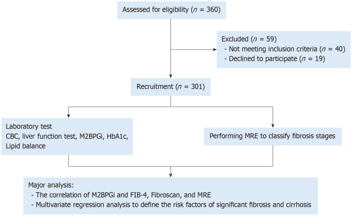
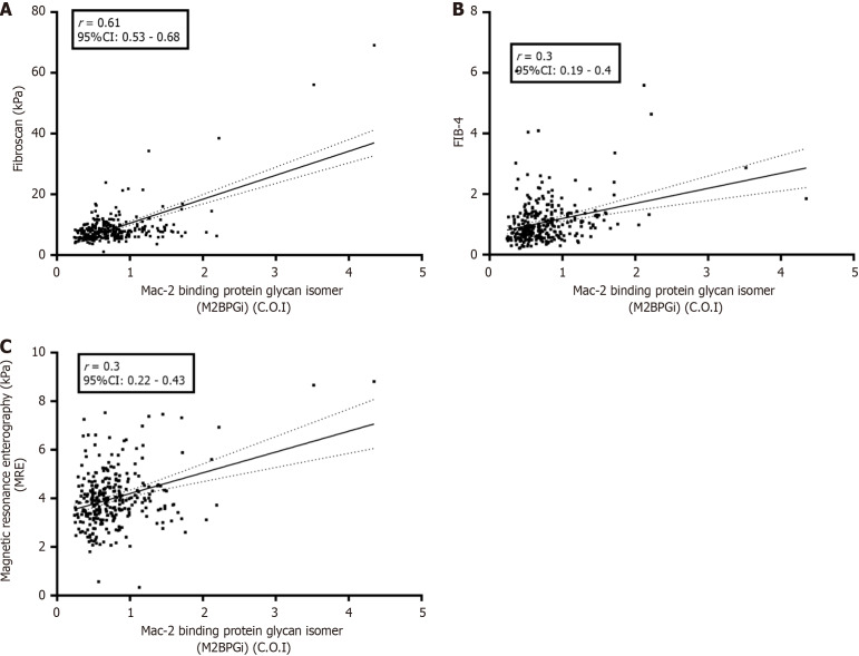
Methods: A total of 301 individuals with ultrasound-confirmed or FibroScan-confirmed diagnosis of fatty liver were enrolled in the study. The participants were stratified according to fibrosis stage, measured via magnetic resonance elastography. M2BPGi, Fibrosis-4 (FIB-4) Index score, and routine parameters of liver function were assessed to statistically investigate the correlation of M2BPGi levels in various fibrosis stages and to identify risk factors associated with fibrosis severity.
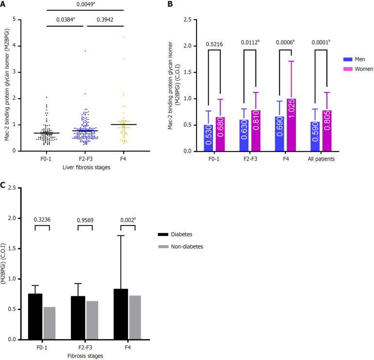
Results: M2BPGi levels positively correlated with fibrosis stages, with cut-off indexes of 0.57 for F0-1, 0.68 for F2-3, and 0.78 for F4. M2BPGi levels in the F0-1 group were significantly different from those in both the F2-3 group (P = 0.038) and the F4 group (P = 0.0051); the F2-3 and F4 groups did not show a significant difference (P = 0.39). Females exhibited significantly higher M2BPGi levels than males for all fibrosis stages, particularly in the F2-3 group (P = 0.01) and F4 group (P = 0.0006). In the F4 (cirrhosis) group, individuals with diabetes had significantly higher M2BPGi levels than those without. M2BPGi, hemoglobin A1c, and FIB-4 score were identified as independent risk factors for greater fibrosis and cirrhosis.
Conclusion: M2BPGi levels varied significantly throughout fibrosis progression, from early MASLD to cirrhosis, with sex correlation. M2BPGi holds promise as an early biomarker for fibrosis characterization in MASLD adult patient populations.

 求助内容:
求助内容: 应助结果提醒方式:


