Liang Wu, Carla C Baan, Derek Reijerkerk, Daan Nieboer, Thierry P P van den Bosch, Dennis A Hesselink, Karin Boer
{"title":"肾移植后急性排斥反应中肾源性尿细胞外囊泡增加:一项初步研究。","authors":"Liang Wu, Carla C Baan, Derek Reijerkerk, Daan Nieboer, Thierry P P van den Bosch, Dennis A Hesselink, Karin Boer","doi":"10.1097/TXD.0000000000001796","DOIUrl":null,"url":null,"abstract":"<p><strong>Background: </strong>Urinary extracellular vesicles (uEVs) are nanosized particles primarily excreted by the kidney. Kidney-derived uEVs (kd-uEVs) are promising noninvasive biomarkers for assessing kidney allograft health and diseases such as acute rejection (AR) after kidney transplantation. However, their release dynamics posttransplant are unclear. This pilot study investigates kd-uEV dynamics and their potential to distinguish AR from acute tubular necrosis (ATN) and nonbiopsied controls.</p><p><strong>Methods: </strong>In the discovery cohort, urine samples from 72 donor-recipient pairs were collected pretransplant and on posttransplant days 3, 7, 180, and before for-cause biopsies. A validation cohort included 28 recipients biopsied within the first 2 wk posttransplant. Urine was stained with CD63 (uEV marker) and kidney-specific markers aquaporin 2 (AQP2) or podocalyxin (PODXL). Kd-uEVs were quantified using imaging flow cytometry, and percentages among total CD63<sup>+</sup> uEVs were calculated to adjust for urine dilution.</p><p><strong>Results: </strong>The percentage of kd-uEVs was lower in pretransplant recipients (AQP2<sup>+</sup>: 1.1% [Q1-Q3, 0.3%-1.7%]; PODXL<sup>+</sup>: 1.5% [Q1-Q3, 0.9%-2.8%]) compared with donors (AQP2<sup>+</sup>: 4.7% [Q1-Q3, 0.9%-11.5%], <i>P</i> < 0.001; PODXL<sup>+</sup> 6.4% [Q1-Q3, 1.4%-9.8%], <i>P</i> < 0.01). Recipients' kd-uEVs remained on pretransplant levels on posttransplant day 3 but were higher on day 7 (AQP2<sup>+</sup>: 7.2% [Q1-Q3, 2.6%-17.4%], <i>P</i> < 0.001; PODXL<sup>+</sup>: 10.0% [Q1-Q3, 3.2%-16.3%], <i>P</i> < 0.001) and persisted until day 180. In the initial 2 wk after transplantation, AR cases had higher AQP2<sup>+</sup> kd-uEVs (17.6% [Q1-Q3, 8.6%-32.3%]) than nonbiopsied controls (6.8% [Q1-Q3, 2.1%-11.2%], <i>P</i> < 0.05) and ATN (1.6% [Q1-Q3, 0.5%-6.4%], <i>P</i> < 0.01), with similar observations for PODXL<sup>+</sup> kd-uEVs. This difference between early AR and ATN was validated in the validation cohort.</p><p><strong>Conclusions: </strong>Kd-uEV release is prominent from day 7 posttransplant. Elevated kd-uEVs are associated with AR, distinguishing it from ATN and demonstrating their potential as noninvasive biomarkers for early AR diagnosis.</p>","PeriodicalId":23225,"journal":{"name":"Transplantation Direct","volume":"11 5","pages":"e1796"},"PeriodicalIF":1.9000,"publicationDate":"2025-04-10","publicationTypes":"Journal Article","fieldsOfStudy":null,"isOpenAccess":false,"openAccessPdf":"https://www.ncbi.nlm.nih.gov/pmc/articles/PMC12313091/pdf/","citationCount":"0","resultStr":"{\"title\":\"Kidney-derived Urinary Extracellular Vesicles are Increased During Acute Rejection after Kidney Transplantation: A Pilot Study.\",\"authors\":\"Liang Wu, Carla C Baan, Derek Reijerkerk, Daan Nieboer, Thierry P P van den Bosch, Dennis A Hesselink, Karin Boer\",\"doi\":\"10.1097/TXD.0000000000001796\",\"DOIUrl\":null,\"url\":null,\"abstract\":\"<p><strong>Background: </strong>Urinary extracellular vesicles (uEVs) are nanosized particles primarily excreted by the kidney. Kidney-derived uEVs (kd-uEVs) are promising noninvasive biomarkers for assessing kidney allograft health and diseases such as acute rejection (AR) after kidney transplantation. However, their release dynamics posttransplant are unclear. This pilot study investigates kd-uEV dynamics and their potential to distinguish AR from acute tubular necrosis (ATN) and nonbiopsied controls.</p><p><strong>Methods: </strong>In the discovery cohort, urine samples from 72 donor-recipient pairs were collected pretransplant and on posttransplant days 3, 7, 180, and before for-cause biopsies. A validation cohort included 28 recipients biopsied within the first 2 wk posttransplant. Urine was stained with CD63 (uEV marker) and kidney-specific markers aquaporin 2 (AQP2) or podocalyxin (PODXL). Kd-uEVs were quantified using imaging flow cytometry, and percentages among total CD63<sup>+</sup> uEVs were calculated to adjust for urine dilution.</p><p><strong>Results: </strong>The percentage of kd-uEVs was lower in pretransplant recipients (AQP2<sup>+</sup>: 1.1% [Q1-Q3, 0.3%-1.7%]; PODXL<sup>+</sup>: 1.5% [Q1-Q3, 0.9%-2.8%]) compared with donors (AQP2<sup>+</sup>: 4.7% [Q1-Q3, 0.9%-11.5%], <i>P</i> < 0.001; PODXL<sup>+</sup> 6.4% [Q1-Q3, 1.4%-9.8%], <i>P</i> < 0.01). Recipients' kd-uEVs remained on pretransplant levels on posttransplant day 3 but were higher on day 7 (AQP2<sup>+</sup>: 7.2% [Q1-Q3, 2.6%-17.4%], <i>P</i> < 0.001; PODXL<sup>+</sup>: 10.0% [Q1-Q3, 3.2%-16.3%], <i>P</i> < 0.001) and persisted until day 180. In the initial 2 wk after transplantation, AR cases had higher AQP2<sup>+</sup> kd-uEVs (17.6% [Q1-Q3, 8.6%-32.3%]) than nonbiopsied controls (6.8% [Q1-Q3, 2.1%-11.2%], <i>P</i> < 0.05) and ATN (1.6% [Q1-Q3, 0.5%-6.4%], <i>P</i> < 0.01), with similar observations for PODXL<sup>+</sup> kd-uEVs. This difference between early AR and ATN was validated in the validation cohort.</p><p><strong>Conclusions: </strong>Kd-uEV release is prominent from day 7 posttransplant. Elevated kd-uEVs are associated with AR, distinguishing it from ATN and demonstrating their potential as noninvasive biomarkers for early AR diagnosis.</p>\",\"PeriodicalId\":23225,\"journal\":{\"name\":\"Transplantation Direct\",\"volume\":\"11 5\",\"pages\":\"e1796\"},\"PeriodicalIF\":1.9000,\"publicationDate\":\"2025-04-10\",\"publicationTypes\":\"Journal Article\",\"fieldsOfStudy\":null,\"isOpenAccess\":false,\"openAccessPdf\":\"https://www.ncbi.nlm.nih.gov/pmc/articles/PMC12313091/pdf/\",\"citationCount\":\"0\",\"resultStr\":null,\"platform\":\"Semanticscholar\",\"paperid\":null,\"PeriodicalName\":\"Transplantation Direct\",\"FirstCategoryId\":\"1085\",\"ListUrlMain\":\"https://doi.org/10.1097/TXD.0000000000001796\",\"RegionNum\":0,\"RegionCategory\":null,\"ArticlePicture\":[],\"TitleCN\":null,\"AbstractTextCN\":null,\"PMCID\":null,\"EPubDate\":\"2025/5/1 0:00:00\",\"PubModel\":\"eCollection\",\"JCR\":\"Q3\",\"JCRName\":\"TRANSPLANTATION\",\"Score\":null,\"Total\":0}","platform":"Semanticscholar","paperid":null,"PeriodicalName":"Transplantation Direct","FirstCategoryId":"1085","ListUrlMain":"https://doi.org/10.1097/TXD.0000000000001796","RegionNum":0,"RegionCategory":null,"ArticlePicture":[],"TitleCN":null,"AbstractTextCN":null,"PMCID":null,"EPubDate":"2025/5/1 0:00:00","PubModel":"eCollection","JCR":"Q3","JCRName":"TRANSPLANTATION","Score":null,"Total":0}
Kidney-derived Urinary Extracellular Vesicles are Increased During Acute Rejection after Kidney Transplantation: A Pilot Study.
Background: Urinary extracellular vesicles (uEVs) are nanosized particles primarily excreted by the kidney. Kidney-derived uEVs (kd-uEVs) are promising noninvasive biomarkers for assessing kidney allograft health and diseases such as acute rejection (AR) after kidney transplantation. However, their release dynamics posttransplant are unclear. This pilot study investigates kd-uEV dynamics and their potential to distinguish AR from acute tubular necrosis (ATN) and nonbiopsied controls.
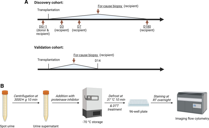
Methods: In the discovery cohort, urine samples from 72 donor-recipient pairs were collected pretransplant and on posttransplant days 3, 7, 180, and before for-cause biopsies. A validation cohort included 28 recipients biopsied within the first 2 wk posttransplant. Urine was stained with CD63 (uEV marker) and kidney-specific markers aquaporin 2 (AQP2) or podocalyxin (PODXL). Kd-uEVs were quantified using imaging flow cytometry, and percentages among total CD63+ uEVs were calculated to adjust for urine dilution.
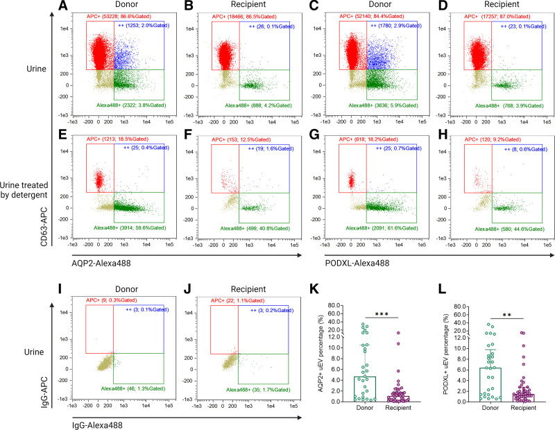
Results: The percentage of kd-uEVs was lower in pretransplant recipients (AQP2+: 1.1% [Q1-Q3, 0.3%-1.7%]; PODXL+: 1.5% [Q1-Q3, 0.9%-2.8%]) compared with donors (AQP2+: 4.7% [Q1-Q3, 0.9%-11.5%], P < 0.001; PODXL+ 6.4% [Q1-Q3, 1.4%-9.8%], P < 0.01). Recipients' kd-uEVs remained on pretransplant levels on posttransplant day 3 but were higher on day 7 (AQP2+: 7.2% [Q1-Q3, 2.6%-17.4%], P < 0.001; PODXL+: 10.0% [Q1-Q3, 3.2%-16.3%], P < 0.001) and persisted until day 180. In the initial 2 wk after transplantation, AR cases had higher AQP2+ kd-uEVs (17.6% [Q1-Q3, 8.6%-32.3%]) than nonbiopsied controls (6.8% [Q1-Q3, 2.1%-11.2%], P < 0.05) and ATN (1.6% [Q1-Q3, 0.5%-6.4%], P < 0.01), with similar observations for PODXL+ kd-uEVs. This difference between early AR and ATN was validated in the validation cohort.
Conclusions: Kd-uEV release is prominent from day 7 posttransplant. Elevated kd-uEVs are associated with AR, distinguishing it from ATN and demonstrating their potential as noninvasive biomarkers for early AR diagnosis.

 求助内容:
求助内容: 应助结果提醒方式:


