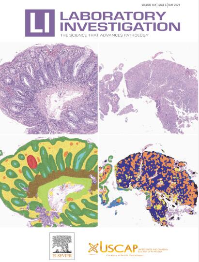
Mohamed Omar , Giuseppe Nicolo’ Fanelli , Fabio Socciarelli , Varun Ullanat , Sreekar Reddy Puchala , James Wen , Alex Chowdhury , Itzel Valencia , Cristian Scatena , Luigi Marchionni , Renato Umeton , Massimo Loda
{"title":"基于抗体的多重图像分析:病理学家的标准分析工作流程和工具。","authors":"Mohamed Omar , Giuseppe Nicolo’ Fanelli , Fabio Socciarelli , Varun Ullanat , Sreekar Reddy Puchala , James Wen , Alex Chowdhury , Itzel Valencia , Cristian Scatena , Luigi Marchionni , Renato Umeton , Massimo Loda","doi":"10.1016/j.labinv.2025.104220","DOIUrl":null,"url":null,"abstract":"<div><div>Conventional histopathology has traditionally been the cornerstone of disease diagnosis, relying on qualitative or semiquantitative visual inspection of tissue sections to detect pathological changes. Singleplex immunohistochemistry (IHC), although effective in detecting specific biomarkers, is often limited by its single-marker focus, which constrains its ability to capture the complexity of the tissue environment. The introduction of multiplexed imaging technologies, such as multiplex IHC and multiplex immunofluorescence, has been transformative, enabling the simultaneous visualization of multiple biomarkers within a single tissue section. These approaches complement morphology with quantitative multimarker data and spatial context, providing a more comprehensive view of cellular interactions and disease mechanisms. However, the rich data from multiplex IHC/multiplex immunofluorescence experiments come with significant analytical challenges, as large multichannel images require comprehensive processing to transform raw imaging data into quantitative and meaningful information. This review focuses on the standard digital image analysis workflow for multiplex imaging in pathology, covering each step from image acquisition and preprocessing to cell segmentation and biomarker quantification. We discuss the common open-source tools that support each step to guide users in selecting appropriate solutions. By outlining an end-to-end pipeline with concrete examples, this review is intended for practicing pathologists and researchers with limited computational expertise. It provides practical guidance and best practices to help integrate multiplex image analysis into routine pathology workflows and translational research, bridging the gap between advanced imaging technology and day-to-day diagnostic practice.</div></div>","PeriodicalId":17930,"journal":{"name":"Laboratory Investigation","volume":"105 10","pages":"Article 104220"},"PeriodicalIF":4.2000,"publicationDate":"2025-07-29","publicationTypes":"Journal Article","fieldsOfStudy":null,"isOpenAccess":false,"openAccessPdf":"","citationCount":"0","resultStr":"{\"title\":\"Antibody-Based Multiplex Image Analysis: Standard Analytical Workflows and Artificial Intelligence Tools for Pathologists\",\"authors\":\"Mohamed Omar , Giuseppe Nicolo’ Fanelli , Fabio Socciarelli , Varun Ullanat , Sreekar Reddy Puchala , James Wen , Alex Chowdhury , Itzel Valencia , Cristian Scatena , Luigi Marchionni , Renato Umeton , Massimo Loda\",\"doi\":\"10.1016/j.labinv.2025.104220\",\"DOIUrl\":null,\"url\":null,\"abstract\":\"<div><div>Conventional histopathology has traditionally been the cornerstone of disease diagnosis, relying on qualitative or semiquantitative visual inspection of tissue sections to detect pathological changes. Singleplex immunohistochemistry (IHC), although effective in detecting specific biomarkers, is often limited by its single-marker focus, which constrains its ability to capture the complexity of the tissue environment. The introduction of multiplexed imaging technologies, such as multiplex IHC and multiplex immunofluorescence, has been transformative, enabling the simultaneous visualization of multiple biomarkers within a single tissue section. These approaches complement morphology with quantitative multimarker data and spatial context, providing a more comprehensive view of cellular interactions and disease mechanisms. However, the rich data from multiplex IHC/multiplex immunofluorescence experiments come with significant analytical challenges, as large multichannel images require comprehensive processing to transform raw imaging data into quantitative and meaningful information. This review focuses on the standard digital image analysis workflow for multiplex imaging in pathology, covering each step from image acquisition and preprocessing to cell segmentation and biomarker quantification. We discuss the common open-source tools that support each step to guide users in selecting appropriate solutions. By outlining an end-to-end pipeline with concrete examples, this review is intended for practicing pathologists and researchers with limited computational expertise. It provides practical guidance and best practices to help integrate multiplex image analysis into routine pathology workflows and translational research, bridging the gap between advanced imaging technology and day-to-day diagnostic practice.</div></div>\",\"PeriodicalId\":17930,\"journal\":{\"name\":\"Laboratory Investigation\",\"volume\":\"105 10\",\"pages\":\"Article 104220\"},\"PeriodicalIF\":4.2000,\"publicationDate\":\"2025-07-29\",\"publicationTypes\":\"Journal Article\",\"fieldsOfStudy\":null,\"isOpenAccess\":false,\"openAccessPdf\":\"\",\"citationCount\":\"0\",\"resultStr\":null,\"platform\":\"Semanticscholar\",\"paperid\":null,\"PeriodicalName\":\"Laboratory Investigation\",\"FirstCategoryId\":\"3\",\"ListUrlMain\":\"https://www.sciencedirect.com/science/article/pii/S0023683725001308\",\"RegionNum\":2,\"RegionCategory\":\"医学\",\"ArticlePicture\":[],\"TitleCN\":null,\"AbstractTextCN\":null,\"PMCID\":null,\"EPubDate\":\"\",\"PubModel\":\"\",\"JCR\":\"Q1\",\"JCRName\":\"MEDICINE, RESEARCH & EXPERIMENTAL\",\"Score\":null,\"Total\":0}","platform":"Semanticscholar","paperid":null,"PeriodicalName":"Laboratory Investigation","FirstCategoryId":"3","ListUrlMain":"https://www.sciencedirect.com/science/article/pii/S0023683725001308","RegionNum":2,"RegionCategory":"医学","ArticlePicture":[],"TitleCN":null,"AbstractTextCN":null,"PMCID":null,"EPubDate":"","PubModel":"","JCR":"Q1","JCRName":"MEDICINE, RESEARCH & EXPERIMENTAL","Score":null,"Total":0}
Antibody-Based Multiplex Image Analysis: Standard Analytical Workflows and Artificial Intelligence Tools for Pathologists
Conventional histopathology has traditionally been the cornerstone of disease diagnosis, relying on qualitative or semiquantitative visual inspection of tissue sections to detect pathological changes. Singleplex immunohistochemistry (IHC), although effective in detecting specific biomarkers, is often limited by its single-marker focus, which constrains its ability to capture the complexity of the tissue environment. The introduction of multiplexed imaging technologies, such as multiplex IHC and multiplex immunofluorescence, has been transformative, enabling the simultaneous visualization of multiple biomarkers within a single tissue section. These approaches complement morphology with quantitative multimarker data and spatial context, providing a more comprehensive view of cellular interactions and disease mechanisms. However, the rich data from multiplex IHC/multiplex immunofluorescence experiments come with significant analytical challenges, as large multichannel images require comprehensive processing to transform raw imaging data into quantitative and meaningful information. This review focuses on the standard digital image analysis workflow for multiplex imaging in pathology, covering each step from image acquisition and preprocessing to cell segmentation and biomarker quantification. We discuss the common open-source tools that support each step to guide users in selecting appropriate solutions. By outlining an end-to-end pipeline with concrete examples, this review is intended for practicing pathologists and researchers with limited computational expertise. It provides practical guidance and best practices to help integrate multiplex image analysis into routine pathology workflows and translational research, bridging the gap between advanced imaging technology and day-to-day diagnostic practice.
期刊介绍:
Laboratory Investigation is an international journal owned by the United States and Canadian Academy of Pathology. Laboratory Investigation offers prompt publication of high-quality original research in all biomedical disciplines relating to the understanding of human disease and the application of new methods to the diagnosis of disease. Both human and experimental studies are welcome.