Christin Schifani, John A. E. Anderson, Arash Nazeri, Aristotle N. Voineskos, the Alzheimer's Disease Neuroimaging Initiative
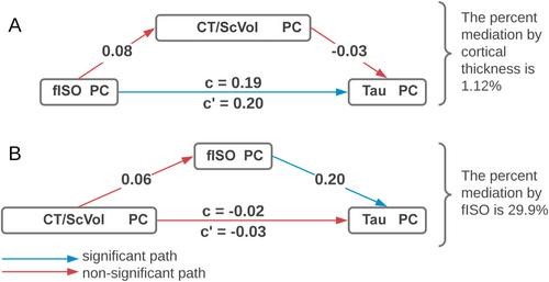
{"title":"体内皮层微结构:与老年人脑损伤和认知障碍的关系","authors":"Christin Schifani, John A. E. Anderson, Arash Nazeri, Aristotle N. Voineskos, the Alzheimer's Disease Neuroimaging Initiative","doi":"10.1111/jnc.70167","DOIUrl":null,"url":null,"abstract":"<p>Positron Emission Tomography (PET) of tau is considered “the” indicator of Alzheimer's pathology. However, non-PET proxies would be helpful for wider accessibility. We used Neurite Orientation Dispersion and Density Imaging (NODDI)-derived indices (i.e., orientation dispersion [ODI], neurite density [NDI], and free-water [fISO]) to determine relationships of gray matter microstructure with tau and cognitive impairment. We assessed the fit between NODDI indices, cortical thickness/subcortical volume (CT/ScVol), and tau via multiple factor analysis (MFA) using data from 80 participants from the ADNI-3 dataset with overlapping multishell diffusion-weighted and tau-PET scans ([<sup>18</sup>F]AV-1451); 49 were considered cognitively normal older adults (age ~74 years), 26 were diagnosed with mild cognitive impairment (age ~75 years), and five had Alzheimer's dementia (age ~78 years). fISO and tau shared a large amount of spatial overlap, and both strongly correlated with the first MFA dimension. Macrostructural features (i.e., CT/ScVol) were 7% less related to this first MFA dimension than fISO and 8% less than tau. Subsequent mediation analyses demonstrated that fISO mediated the relationship between CT/ScVol and tau, explaining all of the variance. Our results suggest that microstructural features derived from NODDI such as fISO might be useful adjunct markers to tau, which needs to be confirmed in longitudinal studies. Cortical fISO, rather than macrostructure (i.e., CT/ScVol), may represent tau's impact on the brain (and, by extension, cognition).</p><p>\n \n <figure>\n <div><picture>\n <source></source></picture><p></p>\n </div>\n </figure>\n </p>","PeriodicalId":16527,"journal":{"name":"Journal of Neurochemistry","volume":"169 8","pages":""},"PeriodicalIF":4.0000,"publicationDate":"2025-08-02","publicationTypes":"Journal Article","fieldsOfStudy":null,"isOpenAccess":false,"openAccessPdf":"https://onlinelibrary.wiley.com/doi/epdf/10.1111/jnc.70167","citationCount":"0","resultStr":"{\"title\":\"In Vivo Cortical Microstructure: Relationships With Tauopathy and Cognitive Impairment in the Elderly\",\"authors\":\"Christin Schifani, John A. E. Anderson, Arash Nazeri, Aristotle N. Voineskos, the Alzheimer's Disease Neuroimaging Initiative\",\"doi\":\"10.1111/jnc.70167\",\"DOIUrl\":null,\"url\":null,\"abstract\":\"<p>Positron Emission Tomography (PET) of tau is considered “the” indicator of Alzheimer's pathology. However, non-PET proxies would be helpful for wider accessibility. We used Neurite Orientation Dispersion and Density Imaging (NODDI)-derived indices (i.e., orientation dispersion [ODI], neurite density [NDI], and free-water [fISO]) to determine relationships of gray matter microstructure with tau and cognitive impairment. We assessed the fit between NODDI indices, cortical thickness/subcortical volume (CT/ScVol), and tau via multiple factor analysis (MFA) using data from 80 participants from the ADNI-3 dataset with overlapping multishell diffusion-weighted and tau-PET scans ([<sup>18</sup>F]AV-1451); 49 were considered cognitively normal older adults (age ~74 years), 26 were diagnosed with mild cognitive impairment (age ~75 years), and five had Alzheimer's dementia (age ~78 years). fISO and tau shared a large amount of spatial overlap, and both strongly correlated with the first MFA dimension. Macrostructural features (i.e., CT/ScVol) were 7% less related to this first MFA dimension than fISO and 8% less than tau. Subsequent mediation analyses demonstrated that fISO mediated the relationship between CT/ScVol and tau, explaining all of the variance. Our results suggest that microstructural features derived from NODDI such as fISO might be useful adjunct markers to tau, which needs to be confirmed in longitudinal studies. Cortical fISO, rather than macrostructure (i.e., CT/ScVol), may represent tau's impact on the brain (and, by extension, cognition).</p><p>\\n \\n <figure>\\n <div><picture>\\n <source></source></picture><p></p>\\n </div>\\n </figure>\\n </p>\",\"PeriodicalId\":16527,\"journal\":{\"name\":\"Journal of Neurochemistry\",\"volume\":\"169 8\",\"pages\":\"\"},\"PeriodicalIF\":4.0000,\"publicationDate\":\"2025-08-02\",\"publicationTypes\":\"Journal Article\",\"fieldsOfStudy\":null,\"isOpenAccess\":false,\"openAccessPdf\":\"https://onlinelibrary.wiley.com/doi/epdf/10.1111/jnc.70167\",\"citationCount\":\"0\",\"resultStr\":null,\"platform\":\"Semanticscholar\",\"paperid\":null,\"PeriodicalName\":\"Journal of Neurochemistry\",\"FirstCategoryId\":\"3\",\"ListUrlMain\":\"https://onlinelibrary.wiley.com/doi/10.1111/jnc.70167\",\"RegionNum\":3,\"RegionCategory\":\"医学\",\"ArticlePicture\":[],\"TitleCN\":null,\"AbstractTextCN\":null,\"PMCID\":null,\"EPubDate\":\"\",\"PubModel\":\"\",\"JCR\":\"Q2\",\"JCRName\":\"BIOCHEMISTRY & MOLECULAR BIOLOGY\",\"Score\":null,\"Total\":0}","platform":"Semanticscholar","paperid":null,"PeriodicalName":"Journal of Neurochemistry","FirstCategoryId":"3","ListUrlMain":"https://onlinelibrary.wiley.com/doi/10.1111/jnc.70167","RegionNum":3,"RegionCategory":"医学","ArticlePicture":[],"TitleCN":null,"AbstractTextCN":null,"PMCID":null,"EPubDate":"","PubModel":"","JCR":"Q2","JCRName":"BIOCHEMISTRY & MOLECULAR BIOLOGY","Score":null,"Total":0}
In Vivo Cortical Microstructure: Relationships With Tauopathy and Cognitive Impairment in the Elderly
Positron Emission Tomography (PET) of tau is considered “the” indicator of Alzheimer's pathology. However, non-PET proxies would be helpful for wider accessibility. We used Neurite Orientation Dispersion and Density Imaging (NODDI)-derived indices (i.e., orientation dispersion [ODI], neurite density [NDI], and free-water [fISO]) to determine relationships of gray matter microstructure with tau and cognitive impairment. We assessed the fit between NODDI indices, cortical thickness/subcortical volume (CT/ScVol), and tau via multiple factor analysis (MFA) using data from 80 participants from the ADNI-3 dataset with overlapping multishell diffusion-weighted and tau-PET scans ([18F]AV-1451); 49 were considered cognitively normal older adults (age ~74 years), 26 were diagnosed with mild cognitive impairment (age ~75 years), and five had Alzheimer's dementia (age ~78 years). fISO and tau shared a large amount of spatial overlap, and both strongly correlated with the first MFA dimension. Macrostructural features (i.e., CT/ScVol) were 7% less related to this first MFA dimension than fISO and 8% less than tau. Subsequent mediation analyses demonstrated that fISO mediated the relationship between CT/ScVol and tau, explaining all of the variance. Our results suggest that microstructural features derived from NODDI such as fISO might be useful adjunct markers to tau, which needs to be confirmed in longitudinal studies. Cortical fISO, rather than macrostructure (i.e., CT/ScVol), may represent tau's impact on the brain (and, by extension, cognition).
期刊介绍:
Journal of Neurochemistry focuses on molecular, cellular and biochemical aspects of the nervous system, the pathogenesis of neurological disorders and the development of disease specific biomarkers. It is devoted to the prompt publication of original findings of the highest scientific priority and value that provide novel mechanistic insights, represent a clear advance over previous studies and have the potential to generate exciting future research.
 求助内容:
求助内容: 应助结果提醒方式:


