{"title":"用SDS-PAGE和荧光显微镜定量测定halotag标记蛋白的细胞内分布。","authors":"Julia Shangguan, Ronald S Rock","doi":"10.21769/BioProtoc.5391","DOIUrl":null,"url":null,"abstract":"<p><p>Counting protein molecules helps reveal the organization of components within cellular structures and the stoichiometries of protein complexes. Existing protein and peptide quantitation methods vary in their complexity. Here, we report a straightforward workflow to measure the absolute number of HaloTag-labeled myosin 10 (Myo10) molecules in U2OS cells. Myo10 is a motor protein that plays a prominent role in cellular protrusion formation. Various biochemical and biological properties of Myo10 are established, but it is not well-defined how many molecules of Myo10 pack into narrow cellular structures called filopodia. We present a workflow for using SDS-PAGE to calibrate Myo10 signal with a reference protein, segmenting epifluorescence microscopy images to map Myo10 intracellular distribution, and interpreting the results to derive biological and functional insights. Our protocol is simple to employ and not only applicable for Myo10 research but also easily adaptable for other biological systems that use HaloTag. Key features • Combining SDS-PAGE densitometry with epifluorescence microscopy to quantitate HaloTag-labeled proteins in cells with readily available equipment. • Details for quantifying protein signal intensity in cellular compartments with semi-automated image segmentation.</p>","PeriodicalId":93907,"journal":{"name":"Bio-protocol","volume":"15 14","pages":"e5391"},"PeriodicalIF":1.1000,"publicationDate":"2025-07-20","publicationTypes":"Journal Article","fieldsOfStudy":null,"isOpenAccess":false,"openAccessPdf":"https://www.ncbi.nlm.nih.gov/pmc/articles/PMC12304454/pdf/","citationCount":"0","resultStr":"{\"title\":\"Quantifying Intracellular Distributions of HaloTag-Labeled Proteins With SDS-PAGE and Epifluorescence Microscopy.\",\"authors\":\"Julia Shangguan, Ronald S Rock\",\"doi\":\"10.21769/BioProtoc.5391\",\"DOIUrl\":null,\"url\":null,\"abstract\":\"<p><p>Counting protein molecules helps reveal the organization of components within cellular structures and the stoichiometries of protein complexes. Existing protein and peptide quantitation methods vary in their complexity. Here, we report a straightforward workflow to measure the absolute number of HaloTag-labeled myosin 10 (Myo10) molecules in U2OS cells. Myo10 is a motor protein that plays a prominent role in cellular protrusion formation. Various biochemical and biological properties of Myo10 are established, but it is not well-defined how many molecules of Myo10 pack into narrow cellular structures called filopodia. We present a workflow for using SDS-PAGE to calibrate Myo10 signal with a reference protein, segmenting epifluorescence microscopy images to map Myo10 intracellular distribution, and interpreting the results to derive biological and functional insights. Our protocol is simple to employ and not only applicable for Myo10 research but also easily adaptable for other biological systems that use HaloTag. Key features • Combining SDS-PAGE densitometry with epifluorescence microscopy to quantitate HaloTag-labeled proteins in cells with readily available equipment. • Details for quantifying protein signal intensity in cellular compartments with semi-automated image segmentation.</p>\",\"PeriodicalId\":93907,\"journal\":{\"name\":\"Bio-protocol\",\"volume\":\"15 14\",\"pages\":\"e5391\"},\"PeriodicalIF\":1.1000,\"publicationDate\":\"2025-07-20\",\"publicationTypes\":\"Journal Article\",\"fieldsOfStudy\":null,\"isOpenAccess\":false,\"openAccessPdf\":\"https://www.ncbi.nlm.nih.gov/pmc/articles/PMC12304454/pdf/\",\"citationCount\":\"0\",\"resultStr\":null,\"platform\":\"Semanticscholar\",\"paperid\":null,\"PeriodicalName\":\"Bio-protocol\",\"FirstCategoryId\":\"1085\",\"ListUrlMain\":\"https://doi.org/10.21769/BioProtoc.5391\",\"RegionNum\":0,\"RegionCategory\":null,\"ArticlePicture\":[],\"TitleCN\":null,\"AbstractTextCN\":null,\"PMCID\":null,\"EPubDate\":\"\",\"PubModel\":\"\",\"JCR\":\"Q3\",\"JCRName\":\"BIOLOGY\",\"Score\":null,\"Total\":0}","platform":"Semanticscholar","paperid":null,"PeriodicalName":"Bio-protocol","FirstCategoryId":"1085","ListUrlMain":"https://doi.org/10.21769/BioProtoc.5391","RegionNum":0,"RegionCategory":null,"ArticlePicture":[],"TitleCN":null,"AbstractTextCN":null,"PMCID":null,"EPubDate":"","PubModel":"","JCR":"Q3","JCRName":"BIOLOGY","Score":null,"Total":0}
Quantifying Intracellular Distributions of HaloTag-Labeled Proteins With SDS-PAGE and Epifluorescence Microscopy.
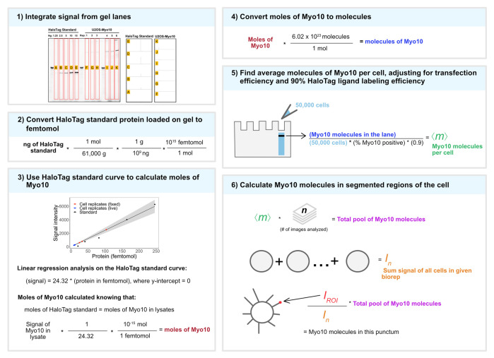
Counting protein molecules helps reveal the organization of components within cellular structures and the stoichiometries of protein complexes. Existing protein and peptide quantitation methods vary in their complexity. Here, we report a straightforward workflow to measure the absolute number of HaloTag-labeled myosin 10 (Myo10) molecules in U2OS cells. Myo10 is a motor protein that plays a prominent role in cellular protrusion formation. Various biochemical and biological properties of Myo10 are established, but it is not well-defined how many molecules of Myo10 pack into narrow cellular structures called filopodia. We present a workflow for using SDS-PAGE to calibrate Myo10 signal with a reference protein, segmenting epifluorescence microscopy images to map Myo10 intracellular distribution, and interpreting the results to derive biological and functional insights. Our protocol is simple to employ and not only applicable for Myo10 research but also easily adaptable for other biological systems that use HaloTag. Key features • Combining SDS-PAGE densitometry with epifluorescence microscopy to quantitate HaloTag-labeled proteins in cells with readily available equipment. • Details for quantifying protein signal intensity in cellular compartments with semi-automated image segmentation.

 求助内容:
求助内容: 应助结果提醒方式:


