Danial Nasiri, Theoni Maragkou, Andreas Raabe, Anna Katharina Krähenbühl, Franca Wagner
{"title":"原发性中枢神经系统粘膜相关淋巴组织淋巴瘤:一个诊断挑战。","authors":"Danial Nasiri, Theoni Maragkou, Andreas Raabe, Anna Katharina Krähenbühl, Franca Wagner","doi":"10.1093/bjrcr/uaaf037","DOIUrl":null,"url":null,"abstract":"<p><p>Primary central nervous system (CNS) mucosa-associated lymphoid tissue (MALT) lymphoma is a rare condition frequently mistaken for meningioma. Since these conditions require distinct treatment approaches, recognizing their imaging characteristics is essential for accurate clinical decision-making. A 69-year-old woman presented with headaches and forehead swelling, prompting MRI of the CNS. Suspecting an intracranial meningioma, the tumour board recommended surgical resection. However, histopathological analysis identified the lesion as a primary CNS MALT lymphoma. Follow-up revealed secondary cutaneous tumour infiltration, leading to a delay in adjuvant radiotherapy. Understanding the differential diagnoses of meningioma is critical for neuroradiologists and neurosurgeons to ensure appropriate treatment planning. This case highlights a misdiagnosis of meningioma that was ultimately identified as a primary CNS MALT lymphoma, emphasizing key imaging and clinical characteristics essential for distinguishing between the most important differential diagnoses of primary CNS MALT lymphoma.</p>","PeriodicalId":45216,"journal":{"name":"BJR Case Reports","volume":"11 4","pages":"uaaf037"},"PeriodicalIF":0.5000,"publicationDate":"2025-07-17","publicationTypes":"Journal Article","fieldsOfStudy":null,"isOpenAccess":false,"openAccessPdf":"https://www.ncbi.nlm.nih.gov/pmc/articles/PMC12308279/pdf/","citationCount":"0","resultStr":"{\"title\":\"Primary central nervous system mucosa-associated lymphoid tissue lymphoma: a diagnostic challenge.\",\"authors\":\"Danial Nasiri, Theoni Maragkou, Andreas Raabe, Anna Katharina Krähenbühl, Franca Wagner\",\"doi\":\"10.1093/bjrcr/uaaf037\",\"DOIUrl\":null,\"url\":null,\"abstract\":\"<p><p>Primary central nervous system (CNS) mucosa-associated lymphoid tissue (MALT) lymphoma is a rare condition frequently mistaken for meningioma. Since these conditions require distinct treatment approaches, recognizing their imaging characteristics is essential for accurate clinical decision-making. A 69-year-old woman presented with headaches and forehead swelling, prompting MRI of the CNS. Suspecting an intracranial meningioma, the tumour board recommended surgical resection. However, histopathological analysis identified the lesion as a primary CNS MALT lymphoma. Follow-up revealed secondary cutaneous tumour infiltration, leading to a delay in adjuvant radiotherapy. Understanding the differential diagnoses of meningioma is critical for neuroradiologists and neurosurgeons to ensure appropriate treatment planning. This case highlights a misdiagnosis of meningioma that was ultimately identified as a primary CNS MALT lymphoma, emphasizing key imaging and clinical characteristics essential for distinguishing between the most important differential diagnoses of primary CNS MALT lymphoma.</p>\",\"PeriodicalId\":45216,\"journal\":{\"name\":\"BJR Case Reports\",\"volume\":\"11 4\",\"pages\":\"uaaf037\"},\"PeriodicalIF\":0.5000,\"publicationDate\":\"2025-07-17\",\"publicationTypes\":\"Journal Article\",\"fieldsOfStudy\":null,\"isOpenAccess\":false,\"openAccessPdf\":\"https://www.ncbi.nlm.nih.gov/pmc/articles/PMC12308279/pdf/\",\"citationCount\":\"0\",\"resultStr\":null,\"platform\":\"Semanticscholar\",\"paperid\":null,\"PeriodicalName\":\"BJR Case Reports\",\"FirstCategoryId\":\"1085\",\"ListUrlMain\":\"https://doi.org/10.1093/bjrcr/uaaf037\",\"RegionNum\":0,\"RegionCategory\":null,\"ArticlePicture\":[],\"TitleCN\":null,\"AbstractTextCN\":null,\"PMCID\":null,\"EPubDate\":\"2025/7/1 0:00:00\",\"PubModel\":\"eCollection\",\"JCR\":\"Q4\",\"JCRName\":\"RADIOLOGY, NUCLEAR MEDICINE & MEDICAL IMAGING\",\"Score\":null,\"Total\":0}","platform":"Semanticscholar","paperid":null,"PeriodicalName":"BJR Case Reports","FirstCategoryId":"1085","ListUrlMain":"https://doi.org/10.1093/bjrcr/uaaf037","RegionNum":0,"RegionCategory":null,"ArticlePicture":[],"TitleCN":null,"AbstractTextCN":null,"PMCID":null,"EPubDate":"2025/7/1 0:00:00","PubModel":"eCollection","JCR":"Q4","JCRName":"RADIOLOGY, NUCLEAR MEDICINE & MEDICAL IMAGING","Score":null,"Total":0}
Primary central nervous system mucosa-associated lymphoid tissue lymphoma: a diagnostic challenge.
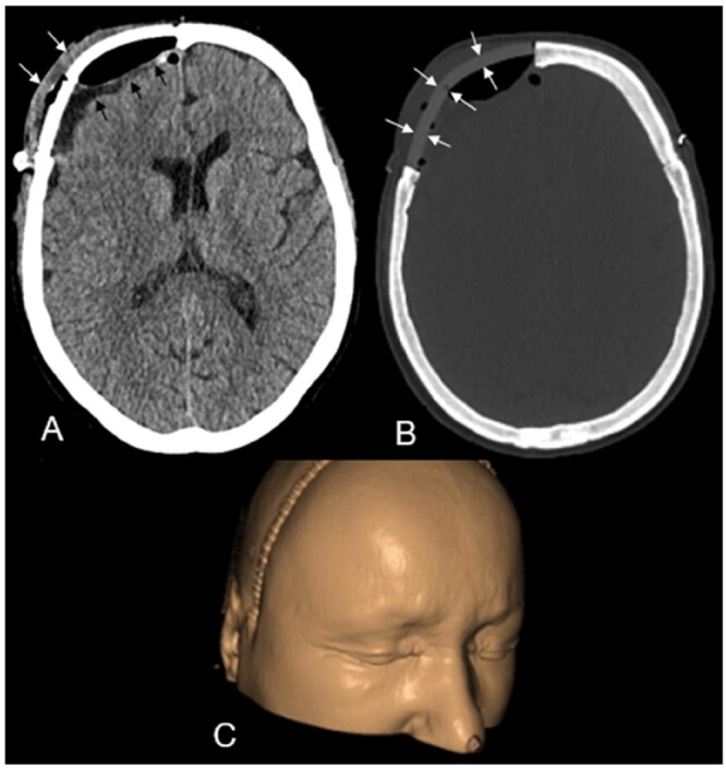
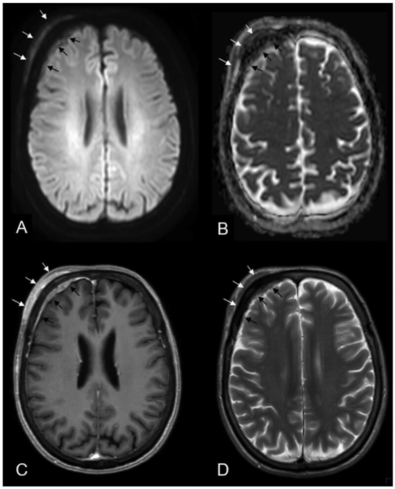
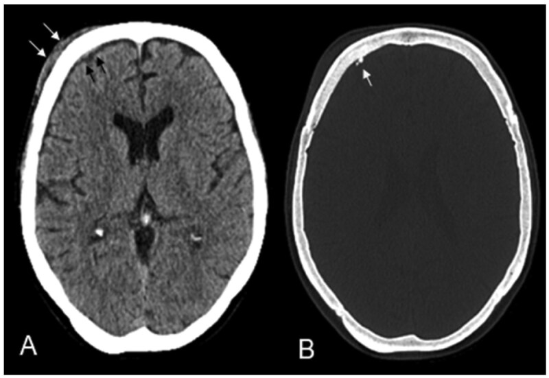
Primary central nervous system (CNS) mucosa-associated lymphoid tissue (MALT) lymphoma is a rare condition frequently mistaken for meningioma. Since these conditions require distinct treatment approaches, recognizing their imaging characteristics is essential for accurate clinical decision-making. A 69-year-old woman presented with headaches and forehead swelling, prompting MRI of the CNS. Suspecting an intracranial meningioma, the tumour board recommended surgical resection. However, histopathological analysis identified the lesion as a primary CNS MALT lymphoma. Follow-up revealed secondary cutaneous tumour infiltration, leading to a delay in adjuvant radiotherapy. Understanding the differential diagnoses of meningioma is critical for neuroradiologists and neurosurgeons to ensure appropriate treatment planning. This case highlights a misdiagnosis of meningioma that was ultimately identified as a primary CNS MALT lymphoma, emphasizing key imaging and clinical characteristics essential for distinguishing between the most important differential diagnoses of primary CNS MALT lymphoma.

 求助内容:
求助内容: 应助结果提醒方式:


