Haijuan Lv, Yu Zhang, Xinyu Wang, Hu Liu, Hongwei Zhao
{"title":"血液透析患者肝铁与脂肪的相关性:定量MRI分析。","authors":"Haijuan Lv, Yu Zhang, Xinyu Wang, Hu Liu, Hongwei Zhao","doi":"10.1016/j.xkme.2025.101035","DOIUrl":null,"url":null,"abstract":"<p><strong>Rationale & objective: </strong>Hepatic iron overload and steatosis are common in hemodialysis patients with anemia who are receiving iron therapy. This study aimed to investigate the relationship between hepatic iron overload and steatosis using magnetic resonance imaging (MRI) while also measuring iron content in extrahepatic organs.</p><p><strong>Study design: </strong>Prospective cohort study.</p><p><strong>Setting & participants: </strong>Fifty-three hemodialysis patients with anemia and a history of iron therapy, along with 45 healthy controls, were recruited from a single center between July 2023 and February 2024.</p><p><strong>Interventions: </strong>All participants underwent 3T MRI with mDIXON-Quant sequences to measure R2∗ and proton density fat fraction (PDFF) values in the liver, pancreas, spleen, and vertebral bone marrow.</p><p><strong>Outcomes: </strong>The primary outcome was the correlation between hepatic R2∗ and PDFF. Secondary analyses evaluated the distribution of iron and fat in extrahepatic organs.</p><p><strong>Results: </strong>Severe hepatic iron overload (R2∗ > 70.1 s<sup>-1</sup>) was observed in 52.8% of patients. Among these patients, hepatic R2∗ was strongly correlated with PDFF (<i>r</i> = 0.67, <i>P</i> < 0.001), whereas a weaker correlation was noted in nonsevere cases (<i>r</i> = 0.16, <i>P</i> = 0.43). Serum ferritin levels were highly correlated with R2∗ in the liver (<i>r</i> = 0.87, <i>P</i> < 0.001), pancreas (<i>r</i> = 0.71, <i>P</i> < 0.001), and spleen (<i>r</i> = 0.78, <i>P</i> < 0.001).</p><p><strong>Limitations: </strong>This single-center study included a relatively small sample size, lacked adjustment for potential confounders, and did not include long-term follow-up.</p><p><strong>Conclusions: </strong>Severe hepatic iron overload is closely associated with elevated liver fat content in hemodialysis patients. These MRI findings may inform more personalized iron therapy strategies by enabling a comprehensive assessment of both hepatic and extrahepatic iron deposition, potentially mitigating treatment-related complications.</p>","PeriodicalId":17885,"journal":{"name":"Kidney Medicine","volume":"7 8","pages":"101035"},"PeriodicalIF":3.4000,"publicationDate":"2025-05-20","publicationTypes":"Journal Article","fieldsOfStudy":null,"isOpenAccess":false,"openAccessPdf":"https://www.ncbi.nlm.nih.gov/pmc/articles/PMC12304958/pdf/","citationCount":"0","resultStr":"{\"title\":\"Correlation Between Hepatic Iron and Fat in Hemodialysis Patients: A Quantitative MRI Analysis.\",\"authors\":\"Haijuan Lv, Yu Zhang, Xinyu Wang, Hu Liu, Hongwei Zhao\",\"doi\":\"10.1016/j.xkme.2025.101035\",\"DOIUrl\":null,\"url\":null,\"abstract\":\"<p><strong>Rationale & objective: </strong>Hepatic iron overload and steatosis are common in hemodialysis patients with anemia who are receiving iron therapy. This study aimed to investigate the relationship between hepatic iron overload and steatosis using magnetic resonance imaging (MRI) while also measuring iron content in extrahepatic organs.</p><p><strong>Study design: </strong>Prospective cohort study.</p><p><strong>Setting & participants: </strong>Fifty-three hemodialysis patients with anemia and a history of iron therapy, along with 45 healthy controls, were recruited from a single center between July 2023 and February 2024.</p><p><strong>Interventions: </strong>All participants underwent 3T MRI with mDIXON-Quant sequences to measure R2∗ and proton density fat fraction (PDFF) values in the liver, pancreas, spleen, and vertebral bone marrow.</p><p><strong>Outcomes: </strong>The primary outcome was the correlation between hepatic R2∗ and PDFF. Secondary analyses evaluated the distribution of iron and fat in extrahepatic organs.</p><p><strong>Results: </strong>Severe hepatic iron overload (R2∗ > 70.1 s<sup>-1</sup>) was observed in 52.8% of patients. Among these patients, hepatic R2∗ was strongly correlated with PDFF (<i>r</i> = 0.67, <i>P</i> < 0.001), whereas a weaker correlation was noted in nonsevere cases (<i>r</i> = 0.16, <i>P</i> = 0.43). Serum ferritin levels were highly correlated with R2∗ in the liver (<i>r</i> = 0.87, <i>P</i> < 0.001), pancreas (<i>r</i> = 0.71, <i>P</i> < 0.001), and spleen (<i>r</i> = 0.78, <i>P</i> < 0.001).</p><p><strong>Limitations: </strong>This single-center study included a relatively small sample size, lacked adjustment for potential confounders, and did not include long-term follow-up.</p><p><strong>Conclusions: </strong>Severe hepatic iron overload is closely associated with elevated liver fat content in hemodialysis patients. These MRI findings may inform more personalized iron therapy strategies by enabling a comprehensive assessment of both hepatic and extrahepatic iron deposition, potentially mitigating treatment-related complications.</p>\",\"PeriodicalId\":17885,\"journal\":{\"name\":\"Kidney Medicine\",\"volume\":\"7 8\",\"pages\":\"101035\"},\"PeriodicalIF\":3.4000,\"publicationDate\":\"2025-05-20\",\"publicationTypes\":\"Journal Article\",\"fieldsOfStudy\":null,\"isOpenAccess\":false,\"openAccessPdf\":\"https://www.ncbi.nlm.nih.gov/pmc/articles/PMC12304958/pdf/\",\"citationCount\":\"0\",\"resultStr\":null,\"platform\":\"Semanticscholar\",\"paperid\":null,\"PeriodicalName\":\"Kidney Medicine\",\"FirstCategoryId\":\"1085\",\"ListUrlMain\":\"https://doi.org/10.1016/j.xkme.2025.101035\",\"RegionNum\":0,\"RegionCategory\":null,\"ArticlePicture\":[],\"TitleCN\":null,\"AbstractTextCN\":null,\"PMCID\":null,\"EPubDate\":\"2025/8/1 0:00:00\",\"PubModel\":\"eCollection\",\"JCR\":\"Q1\",\"JCRName\":\"UROLOGY & NEPHROLOGY\",\"Score\":null,\"Total\":0}","platform":"Semanticscholar","paperid":null,"PeriodicalName":"Kidney Medicine","FirstCategoryId":"1085","ListUrlMain":"https://doi.org/10.1016/j.xkme.2025.101035","RegionNum":0,"RegionCategory":null,"ArticlePicture":[],"TitleCN":null,"AbstractTextCN":null,"PMCID":null,"EPubDate":"2025/8/1 0:00:00","PubModel":"eCollection","JCR":"Q1","JCRName":"UROLOGY & NEPHROLOGY","Score":null,"Total":0}
引用次数: 0
摘要
理由与目的:肝铁超载和脂肪变性在接受铁治疗的血液透析伴贫血患者中很常见。本研究旨在利用磁共振成像(MRI)研究肝铁超载与脂肪变性之间的关系,同时测量肝外器官的铁含量。研究设计:前瞻性队列研究。环境和参与者:在2023年7月至2024年2月期间,从单一中心招募了53名贫血和铁治疗史的血液透析患者,以及45名健康对照者。干预措施:所有参与者都接受了3T MRI和mDIXON-Quant序列来测量肝脏、胰腺、脾脏和脊椎骨髓中的R2 *和质子密度脂肪分数(PDFF)值。结果:主要结果是肝脏R2 *和PDFF之间的相关性。二级分析评估铁和脂肪在肝外器官的分布。结果:52.8%的患者出现严重肝铁超载(R2 * > 70.1 s-1)。在这些患者中,肝脏R2 *与PDFF密切相关(r = 0.67, P r = 0.16, P = 0.43)。血清铁蛋白水平与肝脏中的R2 *高度相关(r = 0.87, P = 0.71, P = 0.78, P)。局限性:该单中心研究样本量相对较小,缺乏对潜在混杂因素的调整,且未包括长期随访。结论:重度肝铁超载与血液透析患者肝脂肪含量升高密切相关。这些MRI结果可以通过全面评估肝脏和肝外铁沉积来提供更个性化的铁治疗策略,潜在地减轻治疗相关的并发症。
Correlation Between Hepatic Iron and Fat in Hemodialysis Patients: A Quantitative MRI Analysis.
Rationale & objective: Hepatic iron overload and steatosis are common in hemodialysis patients with anemia who are receiving iron therapy. This study aimed to investigate the relationship between hepatic iron overload and steatosis using magnetic resonance imaging (MRI) while also measuring iron content in extrahepatic organs.
Study design: Prospective cohort study.
Setting & participants: Fifty-three hemodialysis patients with anemia and a history of iron therapy, along with 45 healthy controls, were recruited from a single center between July 2023 and February 2024.
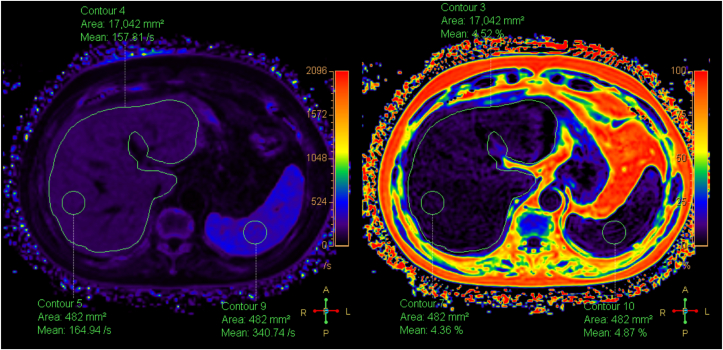
Interventions: All participants underwent 3T MRI with mDIXON-Quant sequences to measure R2∗ and proton density fat fraction (PDFF) values in the liver, pancreas, spleen, and vertebral bone marrow.
Outcomes: The primary outcome was the correlation between hepatic R2∗ and PDFF. Secondary analyses evaluated the distribution of iron and fat in extrahepatic organs.
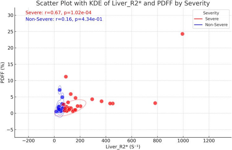
Results: Severe hepatic iron overload (R2∗ > 70.1 s-1) was observed in 52.8% of patients. Among these patients, hepatic R2∗ was strongly correlated with PDFF (r = 0.67, P < 0.001), whereas a weaker correlation was noted in nonsevere cases (r = 0.16, P = 0.43). Serum ferritin levels were highly correlated with R2∗ in the liver (r = 0.87, P < 0.001), pancreas (r = 0.71, P < 0.001), and spleen (r = 0.78, P < 0.001).
Limitations: This single-center study included a relatively small sample size, lacked adjustment for potential confounders, and did not include long-term follow-up.
Conclusions: Severe hepatic iron overload is closely associated with elevated liver fat content in hemodialysis patients. These MRI findings may inform more personalized iron therapy strategies by enabling a comprehensive assessment of both hepatic and extrahepatic iron deposition, potentially mitigating treatment-related complications.
 求助内容:
求助内容: 应助结果提醒方式:


